

Nonsusceptibility of primate cells to Taura syndrome virus
- PMID: 15663846
- PMCID: PMC3323408
- DOI: 10.3201/eid1012.040419
Nonsusceptibility of primate cells to Taura syndrome virus
Abstract
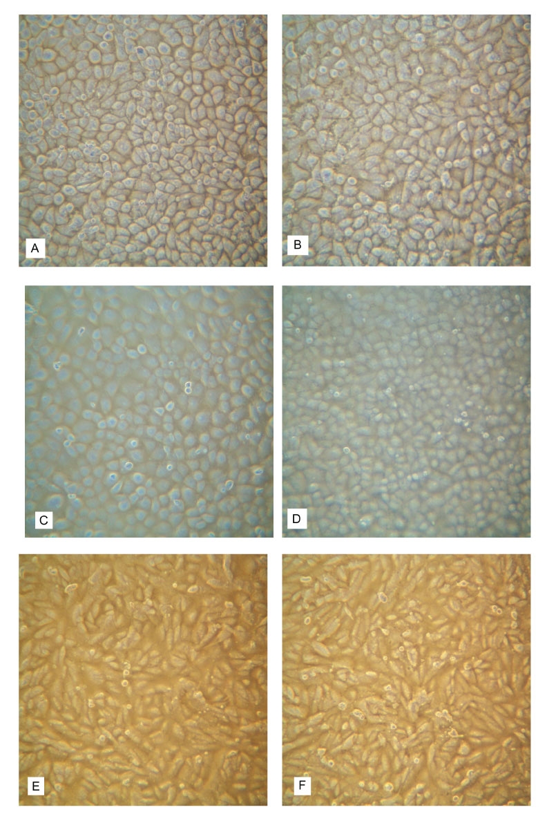
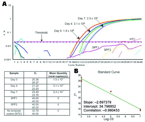
Taura syndrome virus (TSV), a pathogen of penaeid shrimp and member of the family Dicistroviridae, was recently reported to have the ability to infect primate cells. We independently retested this hypothesis. Three lines of primate cells FRhK-4, MA-104, and BGMK, which are highly susceptible to infection by human picornaviruses, were challenged with TSV. Viral replication was assayed by real-time reverse transcription-polymerase chain reaction using cell media samples collected on days 0, 4, and 7 postchallenge. By day 7, genome copy numbers had decreased 25%-99%. No cytopathic effect was observed after 7 days. An in situ hybridization assay, with gene probes specific for detection of TSV, was negative for TSV in challenged cells. The infectivity of residual virus in the cell culture media at day 7 was confirmed by bioassay using TSV-free indicator shrimp (Litopenaeus vannamei). TSV did not infect the primate cells tested, and no evidence of zoonotic potential was found.
Figures
Similar articles
-
Comparison of four Taura syndrome virus (TSV) isolates in oral challenge studies with Litopenaeus vannamei unselected or selected for resistance to TSV.Dis Aquat Organ. 2006 Jul 11;71(1):1-10. doi: 10.3354/dao071001. Dis Aquat Organ. 2006. PMID: 16921995
-
Multiplication of Taura syndrome virus in primary hemocyte culture of shrimp (Penaeus vannamei).J Virol Methods. 2011 Mar;172(1-2):54-9. doi: 10.1016/j.jviromet.2010.12.020. Epub 2010 Dec 28. J Virol Methods. 2011. PMID: 21192980
-
Ultrastructure of the replication site in Taura syndrome virus (TSV)-infected cells.Dis Aquat Organ. 2006 Dec 14;73(2):89-101. doi: 10.3354/dao073089. Dis Aquat Organ. 2006. PMID: 17260828
-
The role of selective breeding and biosecurity in the prevention of disease in penaeid shrimp aquaculture.J Invertebr Pathol. 2012 Jun;110(2):247-50. doi: 10.1016/j.jip.2012.01.013. Epub 2012 Mar 13. J Invertebr Pathol. 2012. PMID: 22434005 Review.
-
Epizootiology, distribution and the impact on international trade of two penaeid shrimp viruses in the Americas.Rev Sci Tech. 1996 Jun;15(2):579-601. doi: 10.20506/rst.15.2.944. Rev Sci Tech. 1996. PMID: 8890382 Review.
Cited by
-
Diverse RNA viruses of arthropod origin in the blood of fruit bats suggest a link between bat and arthropod viromes.Virology. 2019 Feb;528:64-72. doi: 10.1016/j.virol.2018.12.009. Epub 2018 Dec 18. Virology. 2019. PMID: 30576861 Free PMC article.
-
Metagenomic analyses of viruses in stool samples from children with acute flaccid paralysis.J Virol. 2009 May;83(9):4642-51. doi: 10.1128/JVI.02301-08. Epub 2009 Feb 11. J Virol. 2009. PMID: 19211756 Free PMC article.
-
Early qualitative risk assessment of the emerging zoonotic potential of animal diseases.BMJ. 2005 Nov 26;331(7527):1256-60. doi: 10.1136/bmj.331.7527.1256. BMJ. 2005. PMID: 16308389 Free PMC article. Review.
-
Detection of dicistroviruses RNA in blood of febrile Tanzanian children.Emerg Microbes Infect. 2019;8(1):613-623. doi: 10.1080/22221751.2019.1603791. Emerg Microbes Infect. 2019. PMID: 30999808 Free PMC article.
-
Mechanism of translation initiation by Dicistroviridae IGR IRESs.Virology. 2011 Mar 15;411(2):355-61. doi: 10.1016/j.virol.2011.01.005. Epub 2011 Feb 1. Virology. 2011. PMID: 21284991 Free PMC article. Review.
References
Publication types
MeSH terms
LinkOut - more resources
Full Text Sources