. 2005 Feb;73(2):820-7.
doi: 10.1128/IAI.73.2.820-827.2005.

Mannoproteins from Cryptococcus neoformans promote dendritic cell maturation and activation

Affiliations

Mannoproteins from Cryptococcus neoformans promote dendritic cell maturation and activation

Donatella Pietrella et al. Infect Immun. 2005 Feb.

Abstract

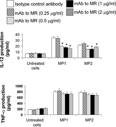
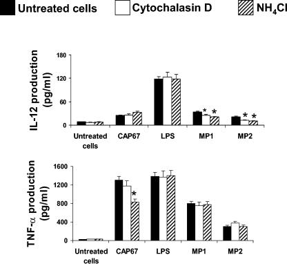
Our previous data show that mannoproteins (MPs) from Cryptococcus neoformans are able to induce protective responses against both C. neoformans and Candida albicans. Here we provide evidence that MPs foster maturation and activation of human dendritic cells (DCs). Maturation was evaluated by the ability of MPs to facilitate expression of costimulatory molecules such as CD40, CD86, CD83, and major histocompatibility complex classes I and II and to inhibit receptors such as CD14, CD16, and CD32. Activation of DCs was measured by the capacity of MPs to promote interleukin-12 and tumor necrosis factor alpha secretion. DC-induced maturation and interleukin-12 induction are largely mediated by engagement of mannose receptors and presume MP internalization and degradation. DC activation leads to IkappaBalpha phosphorylation, which is necessary for nuclear factor kappaB transmigration into the nucleus. MP-loaded DCs are efficient stimulators of T cells and show a remarkable capacity to promote CD4 and CD8 proliferation. In conclusion, we have evidenced a novel regulatory role of MPs that promotes their candidacy as a vaccine against fungi.


Figures

FIG. 1.
FIG. 1.
Cell surface expression of MHC, CD16, CD32, and CS molecules on DCs upon addition of MPs. Immature DCs were treated with different concentrations of MP1, MP2, or LPS (1 μg/ml) for 48 h; stained with FITC-conjugated MAb to CD14, CD16, CD32, CD40, CD83, CD86, MHC class I, or MHC class II; and then examined by flow cytometry. Results are expressed as mean fluorescence intensities (MFIs). The data are representative of one of three independent experiments with similar results.
FIG. 2.
FIG. 2.
Effects of MAb to MRs on CD40 (A), CD86 (B), and CD32 (C) expression induced by MPs. Immature DCs were incubated for 30 min with different concentrations of MAb to MR and then treated with MP1 or MP2 (5 μg/ml) or LPS (1 μg/ml) for 48 h. After incubation, the cells were stained with FITC-conjugated MAb to CD40, CD86, or CD32 and examined by flow cytometry. Results are expressed as mean fluorescence intensities (MFIs). Results represent the mean ± SEM for three separate experiments with cells from three different donors. Double asterisks indicate a P of <0.001, and single asterisks indicate a P of <0.05 (for results with DCs treated with MAb to MR versus results with DCs treated with isotypic control antibody).
FIG. 3.
FIG. 3.
Effect of cytochalasin D and NH4Cl on CD16, CD32, CD40, and CD86 expression induced by MPs. Immature DCs were incubated for 30 min with cytochalasin D (2 μg/ml) or NH4Cl (1 mM) and then challenged with MP1 or MP2 (5 μg/ml) or LPS (1 μg/ml) for 48 h; after incubation, the cells were stained with FITC-conjugated MAb to CD16, CD32, CD40, or CD86 and examined by flow cytometry. Results are expressed as mean fluorescence intensities (MFIs). Results represent the mean ± SEM for four separate experiments with cells from four different donors. Double asterisks indicate a P of <0.001, and single asterisks indicate a P of <0.05 (results with DCs treated with cytochalasin D or NH4Cl versus results with untreated DCs).
FIG. 4.
FIG. 4.
TNF-α (A) and IL-12 (B) production by DCs in response to MPs. A total of 2 × 10⁶ immature DCs/ml were stimulated for 24 h with acapsular C. neoformans CAP67 (4 × 10⁶/ml), MP1 or MP2 (5 μg/ml), or LPS (1 μg/ml). After incubation, supernatants were recovered and then the presence of TNF-α or IL-12 was determined by ELISA. Results represent the mean ± SEM for three separate experiments with cells from three different donors. Double asterisks indicate a P of <0.001, and single asterisks indicate a P of <0.05 (for results with DCs treated with CAP67, LPS, MP1, or MP2 versus results with untreated DCs).
FIG. 5.
FIG. 5.
Effects of MAb to MR on IL-12 and TNF-α production induced by MPs. A total of 2 × 10⁶ immature DCs were incubated for 30 min with different concentrations of MAb to MR and then treated for 24 h with MP1 and MP2 (5 μg/ml). After incubation, supernatants were recovered and then the presence of IL-12 and TNF-α was tested by ELISA. Results, expressed in picograms per milliliter, represent the mean ± SEM for three separate experiments with cells from three different donors. An asterisk indicates a P of <0.05 (for results with DCs treated with MAb to MR versus results with DCs treated with isotype control antibody).
FIG. 6.
FIG. 6.
Effects of cytochalasin D and NH4Cl on IL-12 and TNF-α production induced by MPs. Immature DCs were incubated for 30 min with cytochalasin D (2 μg/ml) or NH4Cl (1 mM) and then challenged with MP1 or MP2 (5 μg/ml), C. neoformans CAP67 (effector-to-target cell ratio, 1:2), or LPS (1 μg/ml) for 24 h. After incubation, supernatants were recovered and then the presence of IL-12 and TNF-α was determined by ELISA. Results, expressed in picograms per milliliter, represent the mean ± SEM for three separate experiments with cells from three different donors. An asterisk indicates a P of <0.05 (for results with DCs treated with cytochalasin D or NH4Cl versus results with untreated DCs).

References

    1. Athamna, A., I. Ofek, Y. Keisari, S. Markowitz, G. G. S. Dutton, and N. Sharon. 1991. Lectinophagocytosis of encapsulated Klebsiella pneumoniae mediated by surface lectins of guinea pig alveolar macrophages and human monocyte-derived macrophages. Infect. Immun. 59:1673-1682. - PMC - PubMed
    1. Bowie, A., and L. A. O'Neill. 2000. Oxidative stress and nuclear factor-κB activation: a reassessment of the evidence in the light of recent discoveries. Biochem. Pharmacol. 59:13-23. - PubMed
    1. Casadevall, A., and J. R. Perfect. 1998. Cryptococcus neoformans, 1st ed. ASM Press, Washington, D.C.
    1. Chaffin, W. L., J. L. López-Ribot, M. Casanova, D. Gozalbo, and J. P. Martínez. 1998. Cell wall and secreted proteins of Candida albicans: identification, function, and expression. Microbiol. Mol. Biol. Rev. 62:130-180. - PMC - PubMed
    1. Chaka, W., A. F. Verheul, V. V. Vaishnav, R. Cherniak, J. Scharringa, J. Verhoef, H. Snippe, and A. I. Hoepelman. 1997. Induction of TNF-alpha in human peripheral blood mononuclear cells by the mannoprotein of Cryptococcus neoformans involves human mannose binding protein. J. Immunol. 159:2979-2985. - PubMed

Publication types

MeSH terms