. 2005 Feb;73(2):878-82.
doi: 10.1128/IAI.73.2.878-882.2005.

Solubility and bioactivity of the Pseudomonas quinolone signal are increased by a Pseudomonas aeruginosa-produced surfactant

Affiliations

Solubility and bioactivity of the Pseudomonas quinolone signal are increased by a Pseudomonas aeruginosa-produced surfactant

M Worth Calfee et al. Infect Immun. 2005 Feb.

Abstract

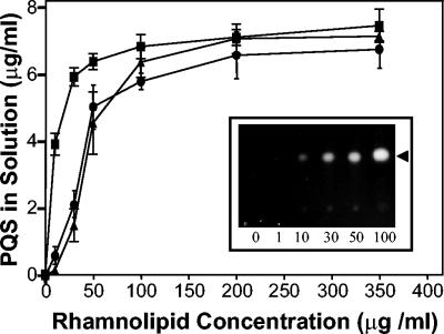
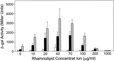
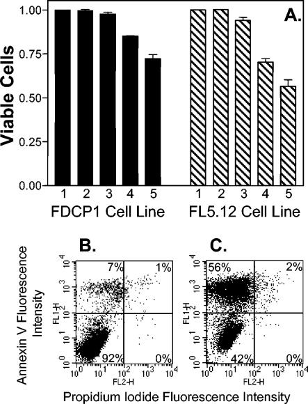
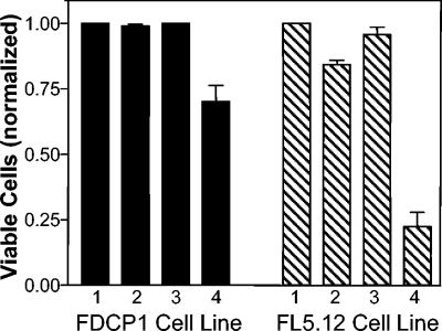
Pseudomonas aeruginosa is a gram-negative bacterium that causes serious infections in immunocompromised individuals and cystic fibrosis patients. This opportunistic pathogen controls many of its virulence factors and cellular functions through the activity of three cell-to-cell signals, N-(3-oxododecanoyl)-L-homoserine lactone, N-butyryl-L-homoserine lactone, and the Pseudomonas quinolone signal (PQS). The activity of these signals is dependent upon their ability to dissolve in and freely diffuse through the aqueous solution in which P. aeruginosa happens to reside. Despite this, our data indicated that PQS was relatively insoluble in aqueous solutions, which led us to postulate that P. aeruginosa could be producing a PQS-solubilizing factor. In this report, we show that the P. aeruginosa-produced biosurfactant rhamnolipid greatly enhances the solubility of PQS in aqueous solutions. The enhanced solubility of PQS led to an increase in PQS bioactivity, as measured by both a gene induction assay and an apoptosis assay. This is the first demonstration of the importance of a bacterial surfactant in the solubilization and bioactivity of a cell-to-cell signal.


Figures

FIG. 1.
FIG. 1.
PQS solubility is low in aqueous solutions. PQS (10 μg) was dried in 13-ml polystyrene tubes, and 0.5 ml of PTSB (lane 1), distilled water (lane 2), or acidified ethyl acetate (lane 3) was added to each tube. Mixtures were vortexed at high speed for 30 min, and aliquots were removed for PQS extraction. Samples were analyzed by TLC as described in Materials and Methods. The figure is a representative TLC plate photographed under UV light. Lane 4 contains 50 ng of synthetic PQS and was included as a standard. The arrowhead indicates PQS.
FIG. 2.
FIG. 2.
Rhamnolipids increase PQS solubility in aqueous solutions. Synthetic PQS (5 μg) and increasing amounts of rhamnolipids were dried in 13-ml polystyrene tubes. Distilled water (circles), phosphate-buffered saline (triangles), or PTSB (squares) (0.5 ml of each) was then added to produce the indicated concentrations of rhamnolipid, and the mixture was incubated at 37°C with vigorous shaking (250 rpm) for 2 h. Aliquots removed from each tube were then organically extracted and analyzed as described in Materials and Methods. The amount of PQS in solution in each mixture was calculated and is presented as the mean ± standard deviation from three separate experiments. The insert is a representative TLC plate photographed under UV light. Numbers within the inset indicate rhamnolipid concentration, and the arrowhead indicates PQS. The PQS solubility assay was performed as in Fig. 1, and the photograph is included to visually show the effects of lower rhamnolipid concentrations on PQS solubility in distilled water.
FIG. 3.
FIG. 3.
Rhamnolipids enhance a PQS bioassay. P. aeruginosa strain PAO-R1(pTS400) (lasR) was grown in the presence of 10 μM (white bars), 20 μM (black bars), or 30 μM (hatched bars) synthetic PQS and increasing amounts of rhamnolipids for 18 h. β-Galactosidase activity was subsequently assayed, and data are presented as the mean in Miller units ± standard deviation of duplicate assays from three separate experiments.
FIG. 4.
FIG. 4.
PQS induces apoptosis. The indicated cell lines were exposed to various physiological concentrations of PQS for 3 days, and the extent of apoptosis was determined by staining with annexin V and propidium iodide. (A) The percentage of viable cells was normalized to 1 for the no-addition control (lane 1) of each cell line. This represents cells which did not stain with either annexin V or propidium iodide. Lane 2 contained 5.2 μl (the maximum amount of solvent used in PQS-containing wells) of dried 1:1 acidified ethyl acetate-acetonitrile as a solvent control. Lanes 3, 4, and 5 contained dried PQS that would produce final concentrations of 1 μM, 5 μM, and 10 μM PQS, respectively, when 2 ml of cell culture was added. Results are presented as the mean ± standard deviation from at least three separate experiments. (B and C) Representative dot plots are included to show the apoptotic shift induced by PQS in the FL5.12 cell line. Panel B shows cells that were grown without PQS (no addition), and panel C shows cells grown in the presence of 10 μM PQS. The percentages given within each panel indicate the percentage of cells in that panel. The lower left panel of each dot plot contains viable cells, as indicated by the lack of staining with either annexin V or propidium iodide. The upper left and upper right panels contain apoptotic cells, which stained with annexin V, and the lower right panels contain dead cells, which stained with propidium iodide only.
FIG. 5.
FIG. 5.
Rhamnolipids enhance PQS-induced apoptosis. PQS and/or rhamnolipids were dried in polystyrene tubes and resuspended in distilled water as described in Material and Methods. The final concentration of rhamnolipid in each tube was 40 μg/ml, and if all PQS became soluble, the final concentration of PQS would be 10 μM. A 200-μl aliquot of suspension (or distilled water as a control) was then added to the indicated cell cultures, which were incubated for 2 days before assaying for apoptosis induction and cell viability as in Fig. 4. Lanes: 1, water; 2, rhamnolipid suspension; 3, PQS suspension; 4, rhamnolipid and PQS suspension. Data were normalized by assigning a value of 1 to the percentage of viable cells found in the distilled-water control (lane 1).

References

    1. Al-Tahhan, R. A., T. R. Sandrin, A. A. Bodour, and R. M. Maier. 2000. Rhamnolipid-induced removal of lipopolysaccharide from Pseudomonas aeruginosa: effect on cell surface properties and interaction with hydrophobic substrates. Appl. Environ. Microbiol. 66:3262-3268. - PMC - PubMed
    1. Bromberg, K. D., A. B. Burgin, and N. Osheroff. 2003. Quinolone action against human topoisomerase IIalpha: stimulation of enzyme-mediated double-stranded DNA cleavage. Biochemistry 42:3393-3398. - PubMed
    1. Calfee, M. W., J. P. Coleman, and E. C. Pesci. 2001. Interference with Pseudomonas quinolone signal synthesis inhibits virulence factor expression by Pseudomonas aeruginosa. Proc. Natl. Acad. Sci. USA 98:11633-11637. - PMC - PubMed
    1. Cao, H., G. Krishnan, B. Goumnerov, J. Tsongalis, R. Tompkins, and L. G. Rahme. 2001. A quorum sensing-associated virulence gene of Pseudomonas aeruginosa encodes a LysR-like transcription regulator with a unique self-regulatory mechanism. Proc. Natl. Acad. Sci. USA 98:14613-14618. - PMC - PubMed
    1. Collier, D. N., L. Anderson, S. L. McKnight, T. L. Noah, M. Knowles, R. Boucher, U. Schwab, P. Gilligan, and E. C. Pesci. 2002. A bacterial cell to cell signal in the lungs of cystic fibrosis patients. FEMS Microbiol. Lett. 215:41-46. - PubMed

Publication types

MeSH terms

LinkOut - more resources