. 2005 Feb;73(2):1052-60.
doi: 10.1128/IAI.73.2.1052-1060.2005.

Effect of capsulation of opportunistic pathogenic bacteria on binding of the pattern recognition molecules mannan-binding lectin, L-ficolin, and H-ficolin

Affiliations

Effect of capsulation of opportunistic pathogenic bacteria on binding of the pattern recognition molecules mannan-binding lectin, L-ficolin, and H-ficolin

Anders Krarup et al. Infect Immun. 2005 Feb.

Abstract

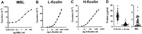
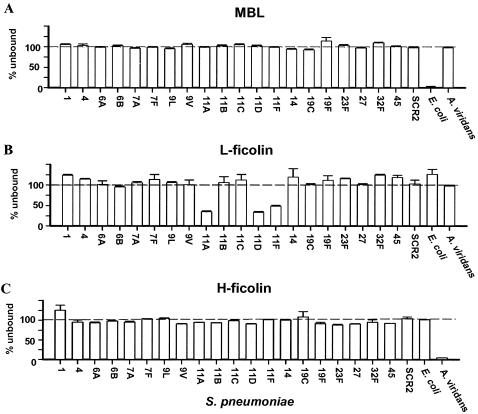
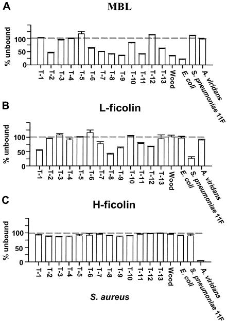
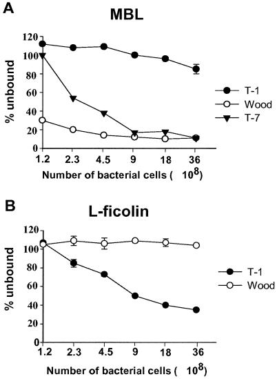
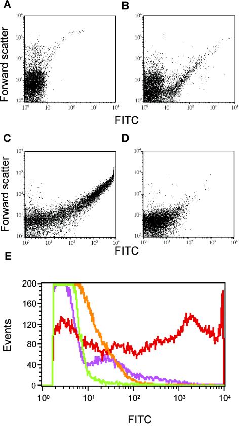
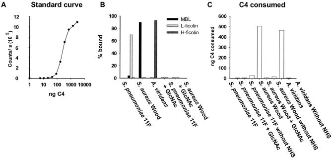
Mannan-binding lectin (MBL), L-ficolin, and H-ficolin are pattern recognition molecules of the innate immune system. We investigated their ability to bind to different serotypes and noncapsulated variants of two gram-positive bacterial species, Streptococcus pneumoniae and Staphylococcus aureus. MBL did not bind to capsulated S. aureus or capsulated S. pneumoniae but did bind to a noncapsulated S. aureus variant (Wood). L-ficolin bound to some capsulated S. aureus serotypes (serotypes 1, 8, 9, 11, and 12) and capsulated S. pneumoniae serotypes (11A, 11D, and 11F) but not to noncapsulated strains. H-ficolin did not bind to any of the S. pneumoniae and S. aureus serotypes included in this study but did bind to one strain of Aerococcus viridans. The concentrations of the three proteins in 97 plasma samples were estimated. The median concentrations were 0.8 mug per ml for MBL, 3.3 mug per ml for L-ficolin, and 18.4 mug per ml for H-ficolin.


Figures

FIG. 1.
FIG. 1.
Assays for the determination of MBL, L-ficolin, and H-ficolin concentrations. Dilutions of standard sera with known concentrations of the respective molecules were applied to wells coated with monoclonal antibodies to MBL (A), L-ficolin (B), and H-ficolin (C). Bound proteins were detected by use of relevant biotinylated monoclonal antibodies. The signals are given as counts per second. Error bars indicate standard deviations. (D) Concentrations of H-ficolin, L-ficolin, and MBL in samples from 97 healthy individuals. Note the different y axis for MBL. Each filled circle represents an individual.
FIG. 2.
FIG. 2.
Binding of MBL, L-ficolin, and H-ficolin to strains of S. pneumoniae and to control strains. MBL, L-ficolin, and H-ficolin in supernatants were measured after incubation of sera and bacteria; binding is seen as a decreased signal. Error bars indicate standard deviations for the individual strains. The results shown are typical of three experiments.
FIG. 3.
FIG. 3.
Binding of MBL, L-ficolin, and H-ficolin to S. aureus. The various serotypes are listed numerically from left to right, with the noncapsulated variant at the right. MBL, L-ficolin, and H-ficolin in supernatants were measured after incubation of sera and bacteria; binding is seen as a decreased signal. Error bars indicate standard deviations for the individual bacteria. The results shown are typical of three experiments.
FIG. 4.
FIG. 4.
Binding of MBL and L-ficolin to dilutions of S. aureus. (A) Amounts of MBL bound to various numbers of serotype T-1, serotype T-7, and strain Wood cells. (B) Amounts of L-ficolin bound to T-1 and Wood. Proteins in supernatants were measured after incubation of sera and bacteria; binding is seen as a decreased signal. Error bars indicate standard deviations.
FIG. 5.
FIG. 5.
Binding of MBL to selected S. aureus serotypes, as analyzed by flow cytometry. Bacteria were incubated with biotinylated rMBL (biotin-rMBL) followed by FITC-labeled streptavidin. (A) Control with bacteria (Wood) and nonbiotinylated rMBL. (B and C) Binding to S. aureus T-1 (B) and S. aureus Wood (C). (D) S. aureus Wood incubated with biotin-rMBL in the presence of 100 mM GlcNAc. The x axis depicts the fluorescence intensity, and the y axis depicts the forward scatter. (E) Compilation of the four flow cytometric histograms, with fluorescence on the x axis and events on the y axis: A, green line; B, purple line; C, red line; D, orange line.
FIG. 6.
FIG. 6.
C4 consumption by MBL, L-ficolin, and H-ficolin complexes bound to bacteria. Bacteria were incubated with sera, washed, and incubated with purified C4, and the amount of residual active C4 was estimated. (A) Standard curve used for quantifying residual C4. (B) Percentages of MBL, L-ficolin, and H-ficolin bound to bacteria. (C) C4 consumed after incubation with bacterium-bound complexes.

References

    1. Akaiwa, M., Y. Yae, R. Sugimoto, S. O. Suzuki, T. Iwaki, K. Izuhara, and N. Hamasaki. 1999. Hakata antigen, a new member of the ficolin/opsonin p35 family, is a novel human lectin secreted into bronchus/alveolus and bile. J. Histochem. Cytochem. 47:777-786. - PubMed
    1. Arizono, T., A. Umeda, and K. Amako. 1991. Distribution of capsular materials on the cell wall surface of strain Smith diffuse of Staphylococcus aureus. J. Bacteriol. 173:4333-4340. - PMC - PubMed
    1. Colley, K. J., M. C. Beranek, and J. U. Baenziger. 1988. Purification and characterization of the core-specific lectin from human serum and liver. Biochem. J. 256:61-68. - PMC - PubMed
    1. Cunnion, K. M., J. C. Lee, and M. M. Frank. 2001. Capsule production and growth phase influence binding of complement to Staphylococcus aureus. Infect. Immun. 69:6796-6803. - PMC - PubMed
    1. Dagnaes-Hansen, F., M. Kilian, and K. Fuursted. 2004. Septicaemia associated with an Aerococcus viridans infection in immunodeficient mice. Lab. Anim. 38:321-325. - PubMed

Publication types

LinkOut - more resources