. 2005 Feb 1;102(5):1614-9.
doi: 10.1073/pnas.0409057102. Epub 2005 Jan 21.

Exposure of acidic residues as a danger signal for recognition of fibrinogen and other macromolecules by integrin alphaXbeta2

Affiliations

Exposure of acidic residues as a danger signal for recognition of fibrinogen and other macromolecules by integrin alphaXbeta2

Thomas Vorup-Jensen et al. Proc Natl Acad Sci U S A. .

Abstract

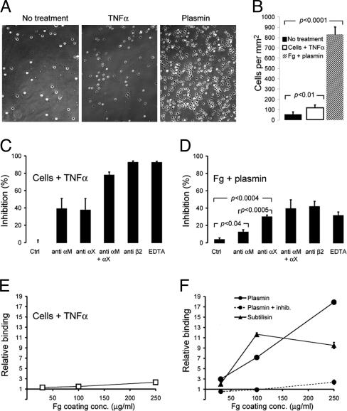
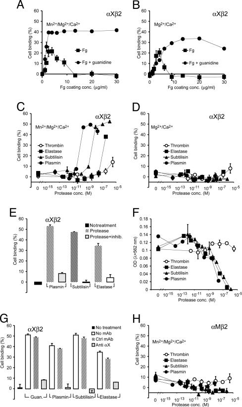
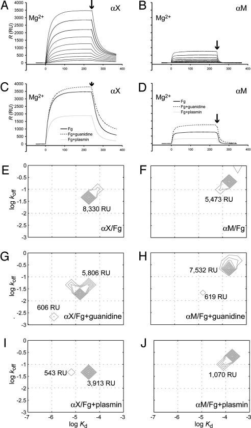
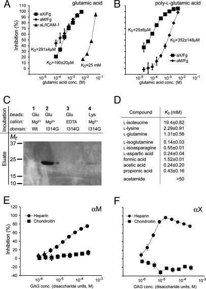
The structural integrity of tissue proteins is damaged in processes ranging from remodeling of the extracellular matrix to destruction by microbial pathogens. Leukocytes play a prominent role in tissue surveillance and repair. However, it remains enigmatic what features of structurally decayed proteins prompt recognition by leukocyte cell-surface receptors. Here, we report that adhesion of human neutrophil granulocytes to fibrinogen is greatly increased by plasmin digestion in a mode where alphaXbeta2 dominates the integrin-dependent binding. The bacterial protease subtilisin also enhances binding by alphaXbeta2. The alphaX ligand binding domain has an unusually high affinity for carboxyl groups, with KD at approximately 100 microM. Our findings implicate enhanced accessibility of negatively charged residues in structurally decayed proteins as a pattern recognition motif for alphaXbeta2 integrin. Comparisons among integrins show relevance of these findings to the large number of ligands recognized by alphaMbeta2 and alphaXbeta2 but not alphaLbeta2. The observations suggest that the pericellular proteolysis at the leading edge of neutrophils not only facilitates passage through the extracellular matrix but also manufactures binding sites for alphaXbeta2.


Figures

Fig. 1.
Fig. 1.
Neutrophil adhesion to immobilized Fg. (A) Micrographs of wells with unstimulated (Left) or TNF-stimulated (Center) human neutrophils binding to Fg, or unstimulated neutrophils binding to plasmin-treated Fg (Right). (B) Average density of neutrophil binding from six different experiments with five different donors (mean value ± SEM). (C) The contribution of integrins to adhesion by TNF-α-stimulated neutrophils was analyzed by addition of an isotypic control Ab, a function blocking anti-αM mAb (CBRM1/29), a function blocking anti-αX mAb (3.9), a combination of these Abs, a function blocking β2 Ab (YCF5.1), or 10 mM EDTA. The percentage of inhibition was calculated from comparison with neutrophil binding in the absence of any addition. (D) Adhesion of unstimulated neutrophils to plasmin-treated Fg. Abs or EDTA were applied as in C. Experiments in AD were carried out in parallel with wells coated with Fg at a concentration of 250 μg/ml. (E and F) The influence of Fg coating on neutrophil adhesion. (E) Binding of TNF-α-stimulated neutrophils. The binding under stimulating conditions at each coating concentration was divided by the binding by unstimulated neutrophils applied in parallel to give relative binding. (F) Binding of unstimulated neutrophils to protease-treated Fg surfaces. In all panels, surfaces were either untreated or treated with 1.3 μM subtilisin, 24 nM plasmin, or 24 nM plasmin in the presence of protease inhibitors. The binding at each coating concentration was divided by the binding in parallel to untreated surfaces to give the relative binding.
Fig. 2.
Fig. 2.
Binding assays with K562 cells expressing recombinant αXβ2 or αMβ2 integrin. For each condition, binding was measured in triplicate wells and stated as mean ± SEM. (A and B) Binding of αXβ2/K562 cells in wells incubated with various concentrations of Fg with or without subsequent treatment by 6 M guanidine. (C and D) Protease induction of αXβ2/K562 cell binding. Wells were coated with 100 μg/ml Fg and treated with plasmin, subtilisin, or neutrophil elastase. All cell adhesion studies were carried out in the presence of 10% (vol/vol) FCS to avoid any effect of residual enzymatic activity in the wells on cellular function. (E) Wells were coated as in C and treated with 1.9 milliunit/ml plasmin (24 nM), 1 milliunit/ml subtilisin (1.3 μM), or 1.6 unit/ml human neutrophil elastase (354 nM) with or without protease inhibitors. (F) Bicinchoninic acid assay of the amount of Fg remaining in the wells after incubation with proteases. (G) Specificity of interaction with αXβ2/K562 cells. A blocking (3.9) or isotypic control Ab to αX was mixed with cells before application to wells either treated with guanidine or proteases. (H) Interaction between proteolyzed Fg and αMβ2/K562 expressing cells was tested in the presence of Mn2+ as for the αXβ2/K562 cells.
Fig. 3.
Fig. 3.
The interaction between Fg and high-affinity αX and αM I domains measured by SPR. The I domains were applied in parallel to flow cells coupled with native (A and B), proteolyzed, or guanidine-treated (C and D)Fginthe presence of 1 mM MgCl2. The affinity for native, proteolyzed, or guanidine-treated Fg for the I domains was measured with a range of 10 concentrations of I domains from 0.28 to 10.6 μM (as shown for native Fg in A and B). For comparison, sensorgrams are shown in C and D from injections of the I domains at the highest applied concentration of 10.6 μM over the surfaces with native, proteolyzed, or guanidine-treated Fg. The end of injection phase is indicated with arrows. (EJ) Two-dimensional off-rate-constant and affinity distribution analyses for heterogeneous surface sites. The calculation was carried out for surfaces with native (E and F), guanidine-treated (G and H), or plasmin-treated (I and J) Fg for measurements either with the αX or αM I domain. The distribution of species at different rate and equilibrium constants is indicated by contour lines, and the total abundance of binding sites in each peak was obtained by integration of the peaks and labeled in response units (RU).
Fig. 4.
Fig. 4.
Interaction between I domain and charged compounds. (A and B) The interaction between the high-affinity αX, αM, and αL I domains and acidic molecules was measured by SPR inhibition assays as described by Karlsson (34). A standard curve for the interaction between various concentrations of each I domain and immobilized ligand (Fg for αX and αM, or ICAM-1 for αL) was established. (A) The I domains were mixed with Glu (inhibitor) in concentrations as indicated, and the response level was converted by use of the standard curve to an estimate of the free amount of I domain (not bound to inhibitor) at each concentration of inhibitor. The amount of inhibitor-bound and free I domain was used to calculate KD. (B) Inhibition of the αX and αM I domains to Fg with poly-l-Glu is shown together with the KD values calculated as in A, with the concentration of inhibitor corresponding to the total Glu concentration. (C) Direct binding of the αX I domain to Glu. Either the wild-type or high-affinity I314G αX I domain was incubated with Glu-coupled beads in the presence of Mg2+ or EDTA as indicated, followed by elution with EDTA, SDS/PAGE, and Coomassie staining. (D) KD values for binding of the αX I domain to various small compounds determined as in A. (E and F) Inhibition of the αM (E) and αX (F) I domain binding to Fg in the presence of heparin or chondroitin sulfate C. Concentrations are given as the molar concentrations of the sulfated disaccharide units of each glycosaminoglycan (GAG) polymer, for comparison to other inhibitors. The Mr of these units was estimated to be ≈500 and 445 for heparin and chondroitin sulfate, respectively.

Similar articles

Cited by

References

    1. Gowen, B. B., Borg, T. K., Ghaffar, A. & Mayer, E. P. (2000) Matrix Biol. 19, 61–71. - PubMed
    1. Gordon, S. (2002) Cell 111, 927–930. - PubMed
    1. Davis, G. E. (1992) Exp. Cell Res. 200, 242–252. - PubMed
    1. Wright, S. D., Weitz, J. I., Huang, A. D., Levin, S. M., Silverstein, S. C. & Loike, J. D. (1988) Proc. Natl. Acad. Sci. USA 85, 7734–7738. - PMC - PubMed
    1. Loike, J. D., Sodeik, B., Cao, L., Leucona, S., Weitz, J. I., Detmers, P. A., Wright, S. D. & Silverstein, S. C. (1991) Proc. Natl. Acad. Sci. USA 88, 1044–1048. - PMC - PubMed

Publication types