. 2005 Feb 1;102(5):1691-6.
doi: 10.1073/pnas.0407661102. Epub 2005 Jan 21.

Transient interference with staphylococcal quorum sensing blocks abscess formation

Affiliations

Transient interference with staphylococcal quorum sensing blocks abscess formation

Jesse S Wright 3rd et al. Proc Natl Acad Sci U S A. .

Abstract

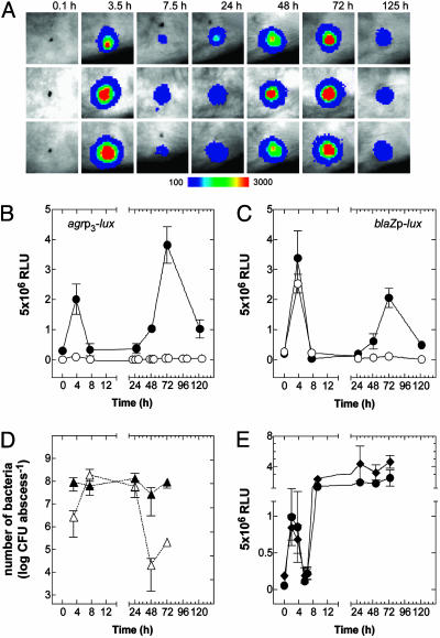
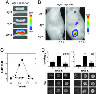
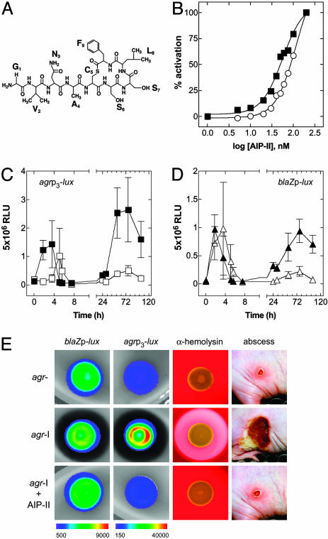
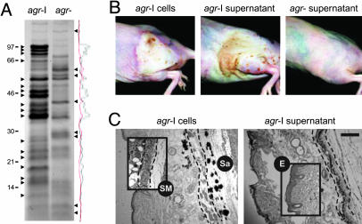
The staphylococcal virulon is controlled largely by the agr locus, a global accessory gene regulator that is autoinduced by a self-coded peptide (AIP) and is therefore a quorum sensor. The agr locus has diverged within and between species, giving rise to AIP variants that inhibit heterologous agr activation, an effect with therapeutic potential against Staphylococcus aureus: a single dose of an inhibitory AIP blocks the formation of an experimental murine abscess. As the AIP is unstable at physiological pH, owing to its essential thiolactone bond, its single-dose efficacy seems paradoxical, which has led us to analyze the in vivo kinetics of agr activation and the consequences of its blockage by a heterologous AIP. Initially, the infecting bacteria grow rapidly, achieving sufficient population density within the first 3 h to activate agr, and then enter a neutrophil-induced metabolic eclipse lasting for 2-3 d, followed by agr reactivation concomitantly with the development of the abscess. The inhibitory AIP prevents agr expression only during its short in vivo lifetime, suggesting that the agr-induced and therefore quorum-dependent synthesis of virulence factors shortly after infection is necessary for the subsequent development of the abscess lesion and bacterial survival. We confirm this finding by showing that a sterile agr+ supernatant causes a sterile abscess similar to the septic abscess caused by live bacteria. These results may provide a biological rationale for regulation of virulence factor expression by quorum sensing rather than by response to specific host signals.


Figures

Fig. 1.
Fig. 1.
Expression of agr during infection. (A) Bioluminescent activity of agr+ (agr-I) bacteria carrying agrp3-lux after s.c. injection in the flank region of SKH-1 mice. Signal intensity is indicated by a pseudocolor scale. (B and C) RLU per lesion were plotted as a function of time for an infecting dose of 108 bacteria carrying either agrp3-lux (B)or blaZp-lux in agr+ (•) and agr (○)(C) backgrounds in groups of three mice. (D) Viable counts of agr+ (▴) and agr (▵) bacteria were enumerated at different points during infection. (E) Activity of agrp3-lux (•) and blaZp-lux (♦) in agr+ bacteria after infection of PMN-depleted mice.
Fig. 2.
Fig. 2.
Autoinduction of agr in vivo.(A) The group-specific AIP-II biosensor strain responds in trans only to an agr-II strain producing its cognate autoinducer, AIP-II, on solid media. (B) A total of 108 agr-I, agr (agr-II::tetM), or agr-II were mixed with equal numbers of the AIP-II biosensor, and a s.c. infection was initiated (n = 3) followed by in vivo imaging. The results with a single mouse represent the series. (C) Bioluminescence generated from 5 × 10⁶ agr-II carrying agrp3-lux (▪) or blaZp-lux (▴) as described in Fig. 1. (D) (Upper) blaZp-lux activities during infection with agr-II and its agr-null derivative. (Lower) The corresponding bacterial colonies obtained by plating identical serial dilutions of the homogenized tissue excised from the site of infection at 0, 3, and 6 h. Each panel shows the bacterial colonies from a single lesion.
Fig. 3.
Fig. 3.
In vivo and in vitro consequences of agr inhibition by a synthetic QS peptide antagonist. (A) Structure of the agr-II autoinducer AIP-II, an agr-I signaling antagonist. (B) Determination of AIP-II half-life. The y axis for the graph indicates the percentage of maximal activation of agrp3-blaZ, and the x axis represents the concentration of AIP-II. The EC50 in PBS was 56.1 nM, and in serum it was 134 nM, indicating that the AIP-II half-life in vivo is <4h.(C and D) The effect of AIP-II on 108 agr-I bacteria carrying agrp3-lux (▪, untreated; □, AIP-II treated) (C) and blaZp-lux (▴, untreated; ▵, AIP-II treated) (D). (E) The effect of AIP-II on bioluminescence of agr-I bacteria carrying blaZp-lux or agrp3-lux when grown on solid media (24 h), on α-hemolysin production when grown on 5% sheep blood agar (24 h), and on lesion size in the murine s.c. model (at 96 h) in comparison with agr.
Fig. 4.
Fig. 4.
Histological comparison of lesions caused by agr+ (agr-I) cells and sterile supernatant. (A) Exoproteins from agr+ and agr supernatants were precipitated with 10% trichloroacetic acid and separated by 10% Tricine SDS/PAGE. After staining with Coomassie brilliant blue and densitometry analysis, several bands were identified that were differentially expressed between the two strains. Sterile culture filtrate was diluted up to 1:4 in PBS, and 0.1 ml was injected without cytodex beads. (B) Gross lesions at 48 h caused by agr+ cells, agr+ supernatant, and agr supernatant. (C) Histological sections of the lesions caused by agr+ cells and agr+ supernatant were stained with hematoxylin and eosin. A Gram stain indicates the dark-stained regions of the lesion caused by agr+ cells that represent clumped staphylococci (Sa). (Insets) For comparison the integrity of the s.c. musculature (SM) (demarcated with dashed lines) and the epidermis (E) in a lesion caused by agr cells is illustrated. (Scale bar: 200 μM.)

References

    1. Shelburne, S. A. & Musser, J. M. (2004) Curr. Opin. Microbiol. 7, 283–289. - PubMed
    1. Novick, R. P. (2003) Mol. Microbiol. 48, 1429–1449. - PubMed
    1. Dunman, P. M., Murphy, E., Haney, S., Palacios, D., Tucker-Kellogg, G., Wu, S., Brown, E. L., Zagursky, R. J., Shlaes, D. & Projan, S. J. (2001) J. Bacteriol. 183, 7341–7353. - PMC - PubMed
    1. Recsei, P., Kreiswirth, B., O'Reilly, M., Schlievert, P., Gruss, A. & Novick, R. P. (1986) Mol. Gen. Genet. 202, 58–61. - PubMed
    1. Abdelnour, A., Arvidson, S., Bremell, T., Ryden, C. & Tarkowski, A. (1993) Infect. Immun. 61, 3879–3885. - PMC - PubMed

Publication types

Substances