

Estrogen receptor alpha and imprinting of the neonatal mouse ventral prostate by estrogen
- PMID: 15665095
- PMCID: PMC547875
- DOI: 10.1073/pnas.0409168102
Estrogen receptor alpha and imprinting of the neonatal mouse ventral prostate by estrogen
Erratum in
- Proc Natl Acad Sci U S A. 2006 May 23;103(21):8298
Abstract
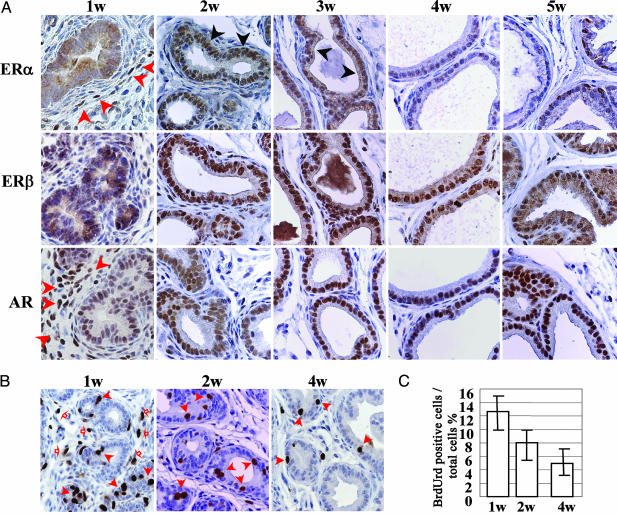
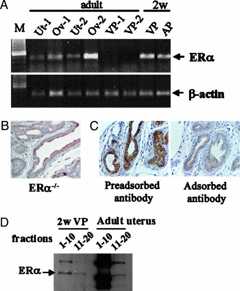
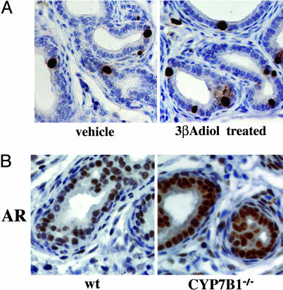
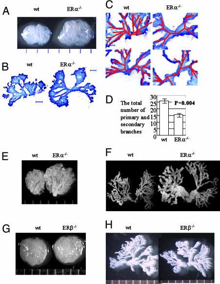
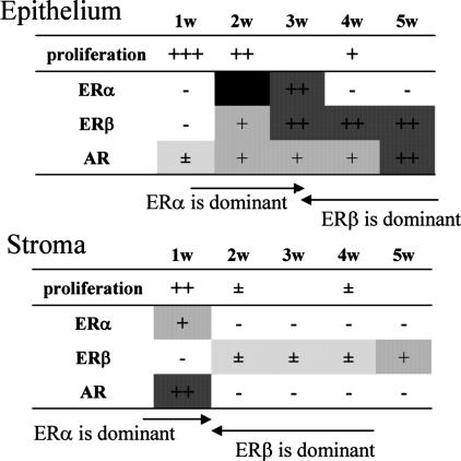
Exposure to estrogen in the neonatal period affects prostatic growth and leads to an increased incidence of prostatic intraepithelial neoplasia in later life. The effects of neonatal estrogen are clearly dependent on estrogen receptor (ER) alpha because they do not occur in ERalpha-knockout mice. Because ERalpha is expressed in the stroma, but not in the epithelium, of the adult ventral prostate, the concept of indirect estrogen effects through stromal signaling has been proposed. Here, we show that during the first 4 weeks of life, there are profound and rapid changes in the ER profile in the mouse ventral prostate. ERalpha is abundant in the stroma during week 1, but by week 2 it is exclusively epithelial, and then by week 4, ERalpha is lost and ERbeta is dominant in the prostatic epithelium. The presence of ERalpha is associated with a high proliferation index, and ERbeta is associated with quiescence. Branching morphogenesis was altered in ERalpha-/-, but not in ERbeta-/-, mice. We conclude that imprinting and branching morphogenesis of the ventral prostate are mediated by estrogen acting directly on epithelial and stromal ERalpha during the first 2 weeks of life.
Figures
Comment in
-
Perinatal imprinting by estrogen and adult prostate disease.Proc Natl Acad Sci U S A. 2005 Feb 1;102(5):1269-70. doi: 10.1073/pnas.0409703102. Epub 2005 Jan 26. Proc Natl Acad Sci U S A. 2005. PMID: 15677320 Free PMC article. No abstract available.
Similar articles
-
Estrogen imprinting of the developing prostate gland is mediated through stromal estrogen receptor alpha: studies with alphaERKO and betaERKO mice.Cancer Res. 2001 Aug 15;61(16):6089-97. Cancer Res. 2001. PMID: 11507058
-
Estrogen receptor-alpha signaling in growth of the ventral prostate: comparison of neonatal growth and postcastration regrowth.Endocrinology. 2008 Sep;149(9):4421-7. doi: 10.1210/en.2007-1413. Epub 2008 Jun 5. Endocrinology. 2008. PMID: 18535112
-
Essential role for estrogen receptor beta in stromal-epithelial regulation of prostatic hyperplasia.Endocrinology. 2007 Feb;148(2):566-74. doi: 10.1210/en.2006-0906. Epub 2006 Oct 26. Endocrinology. 2007. PMID: 17068134
-
The role of Eralpha and ERbeta in the prostate: insights from genetic models and isoform-selective ligands.Ernst Schering Found Symp Proc. 2006;(1):131-47. doi: 10.1007/2789_2006_020. Ernst Schering Found Symp Proc. 2006. PMID: 17824175 Review.
-
Estrogen-regulated development and differentiation of the prostate.Differentiation. 2008 Jul;76(6):660-70. doi: 10.1111/j.1432-0436.2008.00291.x. Epub 2008 Jun 28. Differentiation. 2008. PMID: 18557760 Review.
Cited by
-
Loss of epithelial oestrogen receptor α inhibits oestrogen-stimulated prostate proliferation and squamous metaplasia via in vivo tissue selective knockout models.J Pathol. 2012 Jan;226(1):17-27. doi: 10.1002/path.2949. Epub 2011 Nov 9. J Pathol. 2012. PMID: 22069040 Free PMC article.
-
A recently identified hypothalamic nucleus expressing estrogen receptor alpha.Proc Natl Acad Sci U S A. 2008 Sep 9;105(36):13632-7. doi: 10.1073/pnas.0806503105. Epub 2008 Aug 29. Proc Natl Acad Sci U S A. 2008. PMID: 18757761 Free PMC article.
-
Beta-catenin and estrogen signaling collaborate to drive cyclin D1 expression in developing mouse prostate.Differentiation. 2017 Jan-Feb;93:66-71. doi: 10.1016/j.diff.2016.11.002. Epub 2016 Dec 2. Differentiation. 2017. PMID: 27918915 Free PMC article.
-
Chronic estrogen deficiency causes gastroparesis by altering neuronal nitric oxide synthase function.Dig Dis Sci. 2013 Jun;58(6):1507-15. doi: 10.1007/s10620-013-2610-4. Epub 2013 Mar 17. Dig Dis Sci. 2013. PMID: 23504347 Free PMC article.
-
Imprinting and Reproductive Health: A Toxicological Perspective.Int J Mol Sci. 2023 Nov 21;24(23):16559. doi: 10.3390/ijms242316559. Int J Mol Sci. 2023. PMID: 38068882 Free PMC article. Review.
References
-
- Prins, G. S., Birch, L., Habermann, H., Chang, W. Y., Tebeau, C., Putz, O. & Bieberich, C. (2001) Reprod. Fertil. Dev. 13, 241-252. - PubMed
-
- Strauss, L., Makela, S., Joshi, S., Huhtaniemi, I. & Santti, R. (1998) Mol. Cell. Endocrinol. 144, 83-93. - PubMed
-
- Hayashi, N., Sugimura, Y., Kawamura, J., Donjacour, A. A. & Cunha, G. R. (1991) Biol. Reprod. 45, 308-321. - PubMed
-
- Prins, G. S., Birch, L., Couse, J. F., Choi, I., Katzenellenbogen, B. & Korach, K. S. (2001) Cancer Res. 61, 6089-6097. - PubMed
Publication types
MeSH terms
Substances
LinkOut - more resources
Full Text Sources
Molecular Biology Databases
