. 2005 Feb 1;102(5):1372-7.
doi: 10.1073/pnas.0406347102. Epub 2005 Jan 21.

An intermolecular base triple as the basis of ligand specificity and affinity in the guanine- and adenine-sensing riboswitch RNAs

Affiliations

An intermolecular base triple as the basis of ligand specificity and affinity in the guanine- and adenine-sensing riboswitch RNAs

Jonas Noeske et al. Proc Natl Acad Sci U S A. .

Abstract

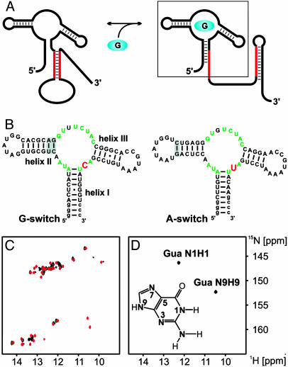
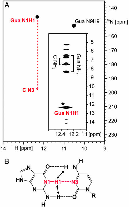
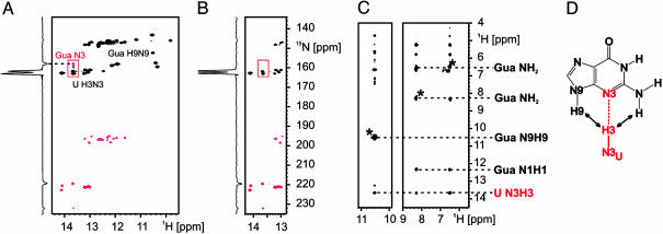
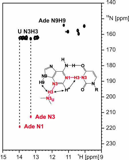
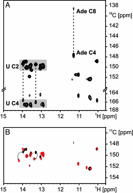
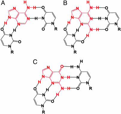
Riboswitches are highly structured RNA elements that control the expression of many bacterial genes by binding directly to small metabolite molecules with high specificity and affinity. In Bacillus subtilis, two classes of riboswitches have been described that discriminate between guanine and adenine despite an extremely high degree of homology both in their primary and secondary structure. We have identified intermolecular base triples between both purine ligands and their respective riboswitch RNAs by NMR spectroscopy. Here, specificity is mediated by the formation of a Watson-Crick base pair between the guanine ligand and a C residue or the adenine ligand and a U residue of the cognate riboswitch RNA, respectively. In addition, a second base-pairing interaction common to both riboswitch purine complexes involves a uridine residue of the RNA and the N3/N9 edge of the purine ligands. This base pairing is mediated by a previously undescribed hydrogen-bonding scheme that contributes to the affinity of the RNA-ligand interaction. The observed intermolecular hydrogen bonds between the purine ligands and the RNA rationalize the previously observed change in specificity upon a C to U mutation in the core of the purine riboswitch RNAs and the differences in the binding affinities for a number of purine analogs.


Figures

Fig. 1.
Fig. 1.
Guanine binding to the aptamer domain of the guanine-responsive riboswitch derived from the 5′ UTR of xpt-pbuX-mRNA in B. subtilis. (A) Conformational change induced by guanine binding to the riboswitch according to ref. . The ligand-binding domain is boxed. (B) Comparison of the secondary structures of the G-switch (Left) and A-switch (Right) RNA ligand-binding domains. Core residues conserved in all purine-binding riboswitch sequences are indicated in green (6, 7). The nucleotide that is conserved as a cytosine in all of the guanine-binding riboswitches and as a uridine in all of the adenine-binding riboswitches is shown in red. Extra residues not found in the original sequence and introduced to facilitate in vitro transcription are denoted in lowercase letters. Mutations in helical regions of the RNAs introduced to stabilize the RNA secondary structure are shaded gray. (C) Overlay of the imino regions of 1H, 15N-HSQC spectra of the uniformly 15N-labeled G-switch RNA in its free form (black) and bound to unlabeled guanine (red) at 283 K. (D) Imino region of an 1H, 15N-HSQC spectrum of 15N-labeled guanine bound to only cytidine-13C, 15N-labeled G-switch RNA. In Inset, the numbering scheme for purines is given with guanine as the example.
Fig. 2.
Fig. 2.
Watson–Crick-type hydrogen bonding between guanine and the G-switch RNA. (A) Imino region of a HNN-COSY experiment using 13C, 15N-C-only labeled RNA and 15N-labeled guanine. The correlation between the guanine H1N1 imino group and a cytosine N3 of the RNA is indicated by a red dashed line. (Inset) Strip for the H1N1 imino group of guanine from a 15N-edited-NOESY experiment. An asterisk denotes the diagonal signal. (B) Hydrogen-bonding scheme for a Watson–Crick G:C base pair with the observed hydrogen bond highlighted and arrows denoting the observed NOE contacts that are typical for a Watson–Crick G:C base pair.
Fig. 3.
Fig. 3.
Hydrogen bonding between the N3/N9 edge of guanine and the G-switch RNA. (A) Imino region of a HNN-COSY experiment using uniformly 15N-labeled RNA and 15N, 13C-labeled guanine. The correlation between the guanine N3 nitrogen and a uridine H3N3 imino group of the RNA is highlighted with a red box. The position of the H9N9 imino group signal of the bound guanine is also indicated. The signal for the guanine H1N1 imino group is overlapped with signals of the RNA. (B) Region of a HNN-COSY experiment by using uniformly 15N-labeled RNA and unlabeled guanine. The correlation between the guanine N3 nitrogen and a uridine H3N3 imino group of the RNA observed in A is absent. One-dimensional slices at a 1H chemical shift of 13.6 ppm are given for the two HNN-COSY experiments in A and B to illustrate the signal-to-noise ratio in the two experiments. (C) Strip for the H9N9 imino group and the amino group of the bound guanine from a 15N-edited-NOESY experiment with the assignment of intermolecular NOEs between the ligand and the U H3N3 imino group of the RNA. The diagonal peaks are indicated with an asterisk. (D) Hydrogen-bonding scheme for the G:U base-pairing interaction with the observed hydrogen bond highlighted in red and arrows denoting the observed intermolecular NOE contacts.
Fig. 4.
Fig. 4.
Hydrogen bonding between adenine and the A-switch RNA. Imino region of a HNN-COSY experiment using only uridine-13C, 15N-labeled RNA and 15N, 13C-labeled adenine. The correlations between two uridine H3N3 imino groups of the A-switch RNA and the N1 and N3 nitrogens of the bound adenine, respectively, are indicated by dashed lines. (Inset) Hydrogen-bonding scheme for the intermolecular base-pairing interactions between adenine and the A-switch RNA with the observed hydrogen bonds highlighted and arrows denoting the observed NOE contacts.
Fig. 5.
Fig. 5.
Carbonyl carbon chemical shifts as indicators of hydrogen bonds. (A) A 2D-H(N)C spectrum of the only uridine 13C, 15N-labeled A-switch RNA in complex with 13C, 15N-labeled adenine. The chemical shift ranges observed for the C2 and C4 carbonyl groups of uridines in Watson–Crick A:U base pairs are shaded in gray. The two uridines involved in intermolecular hydrogen bonds with the ligand (dashed lines) have C2 and C4 chemical shifts close to those for uridines involved in canonical Watson–Crick A:U base pairs. In addition, the correlations between the H9N9 imino group and the C4 and C8 carbons of the bound adenine are indicated. (B) Superimposition of the C2 carbonyl region of 2D-H(N)C spectra of the only uridine 13C, 15N-labeled A-switch RNA in complex with either adenine (black) or 2,6-diaminopurine (red). The chemical shift changes observed for the C2 carbons of the two uridines involved in intermolecular hydrogen bonding with the ligand are indicated by arrows.
Fig. 6.
Fig. 6.
Ligand recognition by an intermolecular base triple in the purine-responsive riboswitches. (A) Intermolecular base-pairing interactions between adenine (red) and two uridines in the adenine-responsive riboswitch. (B) Two additional hydrogen bonds in the complex between 2,6-diaminopurine (red) and the adenine-responsive riboswitch. (C) Intermolecular base-pairing interactions between guanine (red) and nucleotides of the guanine-responsive riboswitch.

References

    1. Barrick, J. E., Corbino, K. A., Winkler, W. C., Nahvi, A., Mandal, M., Collins, J., Lee, M., Roth, A., Sudarsan, N., Jona, I., et al. (2004) Proc. Natl. Acad. Sci. USA 101, 6421-6426. - PMC - PubMed
    1. Sudarsan, N., Barrick, J. E. & Breaker, R. R. (2003) RNA 9, 644-647. - PMC - PubMed
    1. Mandal, M. & Breaker, R. R. (2004) Nat. Rev. Mol. Cell Biol. 5, 451-463. - PubMed
    1. Nudler, E. & Mironov, A. S. (2004) Trends Biochem. Sci. 29, 11-17. - PubMed
    1. Grundy, F. J. & Henkin, T. M. (2004) Curr. Opin. Microbiol. 7, 126-131. - PubMed

Publication types

LinkOut - more resources