. 2005 Feb 1;102(5):1655-60.
doi: 10.1073/pnas.0405488102. Epub 2005 Jan 21.

Attenuation of cardiac remodeling after myocardial infarction by muscle LIM protein-calcineurin signaling at the sarcomeric Z-disc

Affiliations

Attenuation of cardiac remodeling after myocardial infarction by muscle LIM protein-calcineurin signaling at the sarcomeric Z-disc

Joerg Heineke et al. Proc Natl Acad Sci U S A. .

Abstract

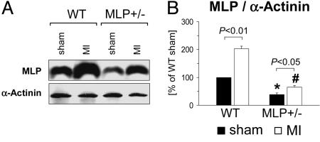
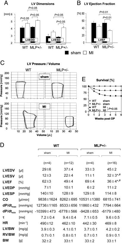
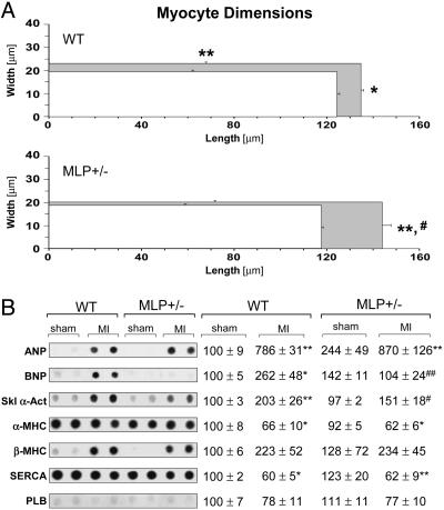
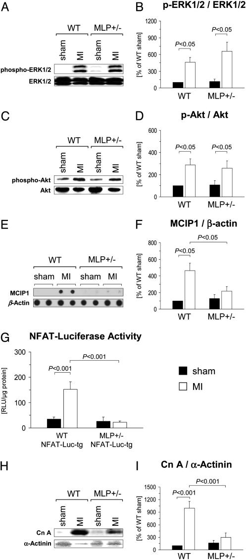
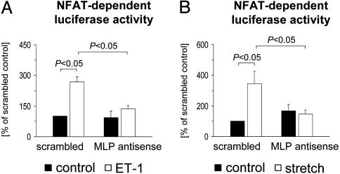
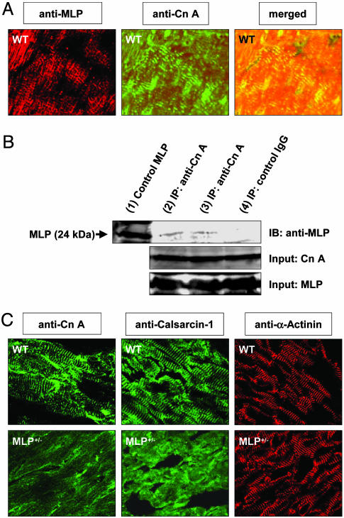
Adverse left ventricular (LV) remodeling after myocardial infarction (MI) is a major cause for heart failure. Molecular modifiers of the remodeling process remain poorly defined. Patients with heart failure after MI have reduced LV expression levels of muscle LIM protein (MLP), a component of the sarcomeric Z-disk that is involved in the integration of stress signals in cardiomyocytes. By using heterozygous MLP mutant (MLP+/-) mice, we explored the role of MLP in post-MI remodeling. LV dimensions and function were similar in sham-operated WT and MLP+/- mice. After MI, however, MLP+/- mice displayed more pronounced LV dilatation and systolic dysfunction and decreased survival compared with WT mice, indicating that reduced MLP levels predispose to adverse LV remodeling. LV dilatation in MLP+/- mice was associated with reduced thickening but enhanced elongation of cardiomyocytes. Activation of the stress-responsive, prohypertrophic calcineurin-nuclear factor of activated T-cells (NFAT) signaling pathway was reduced in MLP+/- mice after MI, as shown by a blunted transcriptional activation of NFAT in cardiomyocytes isolated from MLP+/-/NFAT-luciferase reporter gene transgenic mice. Calcineurin was colocalized with MLP at the Z-disk in WT mice but was displaced from the Z-disk in MLP+/- mice, indicating that MLP is essential for calcineurin anchorage to the Z-disk. In vitro assays in cardiomyocytes with down-regulated MLP confirmed that MLP is required for stress-induced calcineurin-NFAT activation. Our study reveals a link between the stress sensor MLP and the calcineurin-NFAT pathway at the sarcomeric Z-disk in cardiomyocytes and indicates that reduced MLP-calcineurin signaling predisposes to adverse remodeling after MI.


Figures

Fig. 1.
Fig. 1.
Reduced myocardial MLP expression levels in MLP+/– mice. LV myocardial MLP and α-actinin protein expression levels were determined by immunoblotting 6 weeks after sham operation or MI. (A) Typical blots are shown. (B) Data from n = 4–5 mice per group are summarized. *, P < 0.05 vs. sham-operated WT mice; #, P < 0.01 vs. infarcted WT mice.
Fig. 2.
Fig. 2.
Enhanced LV dilatation, reduced systolic function, and increased mortality after MI in MLP+/– mice. (A and B) Echocardiography was performed 6 weeks after sham operation (filled bars) or MI (empty bars). Data are from n = 7 sham-operated mice per genotype, n = 18 infarcted WT mice, and n = 30 infarcted MLP+/– mice. LVEDD and LVESD denote LV end-diastolic and end-systolic diameters, respectively; LVEF, LV ejection fraction. (C and D) Pressure-volume loops were recorded 6 weeks after sham operation or MI. (C) Representative recordings are shown. (D) Data are summarized. LVEDV and LVESV denote LV end-diastolic and endsystolic volumes, respectively; LVEF, LV ejection fraction; LVEDP and LVESP, LV end-systolic and end-diastolic pressures, respectively; CO, cardiac output; dP/dtmax, maximal rate of pressure development; dP/dtmin, maximal rate of pressure decay; τ, monoexponential time constant of LV relaxation; HR, heart rate; LV, LV weight; RV, right ventricular weight; BW, body weight. *, P < 0.05 vs. sham-operated MLP+/– mice; #, P < 0.05 vs. infarcted WT mice. (E) Six-week mortality rates were assessed in 30 WT and 61 MLP+/– mice surviving for at least 48 h after coronary ligation. Sham-operated WT (n = 8) and MLP+/– mice (n = 7) served as controls (survival curves in sham-operated mice are superimposed).
Fig. 3.
Fig. 3.
Cardiomyocyte morphology and gene expression after MI. (A) Cardiomyocyte dimensions were determined 6 weeks after sham operation (white rectangles) or MI (gray rectangles). Approximately 100 myocytes per heart were analyzed in n = 4–5 mice per group. (B) Gene expression levels were determined by mRNA dot blot. Data were normalized to β-actin expression and expressed as [%] of sham-operated WT. (Left) Representative blots are shown (two animals per group). (Right) Data from n = 4–5 sham-operated mice and n = 7–11 infarcted mice per group are presented. *, P < 0.05; **, P < 0.01 vs. sham-operated mice of same genotype; #, P < 0.05; ##, P < 0.01 vs. infarcted WT mice. ANP, atrial natriuretic peptide; SERCA, sarco(endo)plasmic reticulum Ca2+ ATPase-2; PLB, phospholamban.
Fig. 4.
Fig. 4.
Signaling pathway activation in WT and MLP+/– mice after MI. (AD)LV myocardial expression levels of ERK1/2, phospho-ERK1/2, Akt1, and phospho-Akt were determined by immunoblotting 6 weeks after sham operation or MI. (A and C) Typical blots are shown. (B and D) Data from n = 3–5 sham-operated mice (filled bars) and n = 9–10 infarcted mice (empty bars) per group are presented. (E and F) Expression of the exon 4 variant of MCIP1 was determined by mRNA dot-blot analysis and normalized to β-actin expression. (E) Typical blots from two animals per group are shown. (F) Data from n = 3–6 sham-operated mice (filled bars) and n = 7–11 infarcted mice (empty bars) are summarized. (G) NFAT-luciferase reporter activity was measured in cardiomyocytes isolated from MLP+/+(WT)/NFAT-Luc-tg and MLP+/–/NFAT-Luc-tg mice 3 weeks after MI (empty bars) or sham (filled bars) operation. Data from three to five animals per group are shown. (H and I) Calcineurin A and α-actinin protein expression levels were analyzed by immunoblotting. (H) Typical blots are shown. (I) Data from n = 6–7 animals per group (filled bars, sham; empty bars, MI) are summarized.
Fig. 5.
Fig. 5.
MLP is required for NFAT activation in cardiomyocytes. Cultured cardiomyocytes were transfected with an MLP antisense oligonucleotide or a scrambled control oligonucleotide (0.5 μM each). All cells were cotransfected with a luciferase reporter plasmid driven by three NFAT consensus binding sites. Cells were then stimulated for 24 h with 25 nM endothelin-1 (A) or subjected to 24 h of cyclic stretch (B). Data from n = 4–6 experiments are presented.
Fig. 6.
Fig. 6.
MLP is required for calcineurin anchorage to the sarcomeric Z-disk. (A) A LV cryosection obtained from a WT mouse was coimmunostained against MLP (Left) and calcineurin A (Cn A, Center) and analyzed by confocal laser microscopy. (Right) The merged image indicates colocalization of MLP and calcineurin. (B) Immunoprecipitation (IP) of MLP by the anti-calcineurin A antibody (lanes 2 and 3), but not IgG control antibody (lane 4), from WT LV tissue lysates reveals a physical interaction of MLP and calcineurin. A MLP standard was loaded in lane 1. The calcineurin A and MLP IP inputs are shown; IB denotes immunoblotting. (C) Adjacent LV tissue sections from WT and MLP+/– mice were immunostained against calcineurin A, calsarcin-1, and α-actinin and analyzed by confocal laser microscopy. The striated staining pattern of calcineurin A and calsarcin-1 observed in WT mice is lost in MLP+/– mice. By contrast, anti-α-actinin staining reveals prominent striations in WT and MLP+/– mice.

References

    1. Gheorghiade, M. & Bonow, R. O. (1998) Circulation 97, 282–289. - PubMed
    1. Cohn, J. N., Ferrari, R. & Sharpe, N. (2000) J. Am. Coll. Cardiol. 35, 569–582. - PubMed
    1. Mann, D. L. (1999) Circulation 100, 999–1008. - PubMed
    1. Force, T., Michael, A., Kilter, H. & Haq, S. (2002) J. Card. Fail. 8, S351–S358. - PubMed
    1. Zimmer, H. G., Gerdes, A. M., Lortet, S. & Mall, G. (1990) J. Mol. Cell. Cardiol. 22, 1231–1243. - PubMed

Publication types

MeSH terms