

The DNA/RNA-binding protein MSY2 marks specific transcripts for cytoplasmic storage in mouse male germ cells
- PMID: 15665108
- PMCID: PMC547816
- DOI: 10.1073/pnas.0404685102
The DNA/RNA-binding protein MSY2 marks specific transcripts for cytoplasmic storage in mouse male germ cells
Abstract
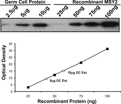
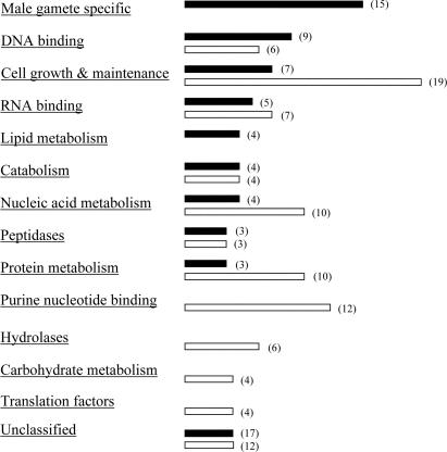
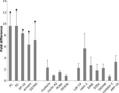
During spermatogenesis, male germ cells temporally synthesize many proteins as they differentiate through meiosis and become spermatozoa. The germ cell Y-box protein, MSY2, constituting approximately 0.7% of total protein in male germ cells, binds to a consensus promoter element, and shows a general lack of RNA-binding specificity. Combining immunoprecipitation and suppressive subtractive hybridization, we identified populations of germ cell mRNAs that are not bound or bound by MSY2. The former population is enriched in cell growth and ubiquitously expressed mRNAs, whereas the latter population is enriched for stored or translationally delayed, male gamete-specific transcripts. Chromatin precipitation assays reveal that most of the MSY2 target mRNAs are transcribed from genes containing the Y-box DNA-binding motif in their promoters. In transgenic mice, mRNAs encoding exogenous GFP are directed or not directed into the MSY2-bound fraction by promoters containing or lacking the Y-box motif, respectively. We propose that MSY2 marks specific mRNAs in the nucleus for cytoplasmic storage, thereby linking transcription and mRNA storage/translational delay in meiotic and postmeiotic male germ cells of the mouse.
Figures
Similar articles
-
In the absence of the mouse DNA/RNA-binding protein MSY2, messenger RNA instability leads to spermatogenic arrest.Biol Reprod. 2007 Jan;76(1):48-54. doi: 10.1095/biolreprod.106.055095. Epub 2006 Oct 11. Biol Reprod. 2007. PMID: 17035640
-
Absence of the DNA-/RNA-binding protein MSY2 results in male and female infertility.Proc Natl Acad Sci U S A. 2005 Apr 19;102(16):5755-60. doi: 10.1073/pnas.0408718102. Epub 2005 Apr 11. Proc Natl Acad Sci U S A. 2005. PMID: 15824319 Free PMC article.
-
Deletion of the DNA/RNA-binding protein MSY2 leads to post-meiotic arrest.Mol Cell Endocrinol. 2006 May 16;250(1-2):20-4. doi: 10.1016/j.mce.2005.12.019. Epub 2006 Jan 18. Mol Cell Endocrinol. 2006. PMID: 16413673 Review.
-
Mammalian male and female germ cells express a germ cell-specific Y-Box protein, MSY2.Biol Reprod. 1998 Nov;59(5):1266-74. doi: 10.1095/biolreprod59.5.1266. Biol Reprod. 1998. PMID: 9780336
-
MSY2 and polypyrimidine tract binding protein 2 stabilize mRNAs in the mammalian testis.Int J Androl. 2008 Sep;31(5):457-61. doi: 10.1111/j.1365-2605.2008.00885.x. Epub 2008 Mar 31. Int J Androl. 2008. PMID: 18380784 Review.
Cited by
-
MIWI associates with translational machinery and PIWI-interacting RNAs (piRNAs) in regulating spermatogenesis.Proc Natl Acad Sci U S A. 2006 Sep 5;103(36):13415-20. doi: 10.1073/pnas.0605506103. Epub 2006 Aug 24. Proc Natl Acad Sci U S A. 2006. PMID: 16938833 Free PMC article.
-
Purification of GidA protein, a novel topoisomerase II inhibitor produced by Streptomyces flavoviridis.World J Microbiol Biotechnol. 2014 Feb;30(2):555-65. doi: 10.1007/s11274-013-1475-1. Epub 2013 Aug 31. World J Microbiol Biotechnol. 2014. PMID: 23996636
-
Basic and clinical genetic studies on male infertility in Iran during 2000-2016: A review.Int J Reprod Biomed. 2018 Mar;16(3):131-148. Int J Reprod Biomed. 2018. PMID: 29766145 Free PMC article. Review.
-
Occurrence and functional significance of the transcriptome in bovine (Bos taurus) spermatozoa.Sci Rep. 2017 Mar 9;7:42392. doi: 10.1038/srep42392. Sci Rep. 2017. PMID: 28276431 Free PMC article.
-
The mouse cytosine-5 RNA methyltransferase NSun2 is a component of the chromatoid body and required for testis differentiation.Mol Cell Biol. 2013 Apr;33(8):1561-70. doi: 10.1128/MCB.01523-12. Epub 2013 Feb 11. Mol Cell Biol. 2013. PMID: 23401851 Free PMC article.
References
Publication types
MeSH terms
Substances
Grants and funding
LinkOut - more resources
Full Text Sources
Molecular Biology Databases
