. 2005;15(1-4):59-68.
doi: 10.1159/000083653.

Regulation of choline deficiency apoptosis by epidermal growth factor in CWSV-1 rat hepatocytes

Affiliations

Regulation of choline deficiency apoptosis by epidermal growth factor in CWSV-1 rat hepatocytes

Craig D Albright et al. Cell Physiol Biochem. 2005.

Abstract

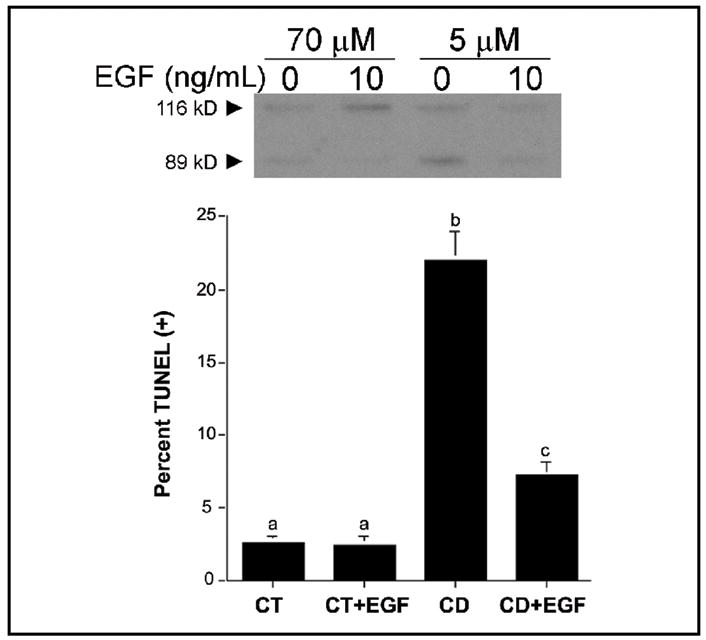
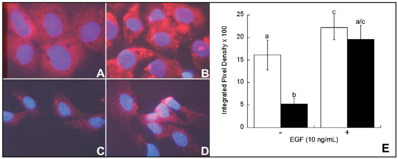
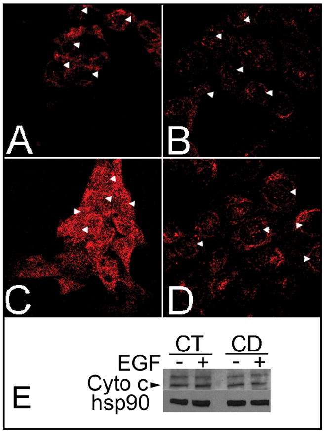
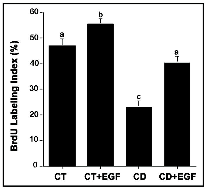
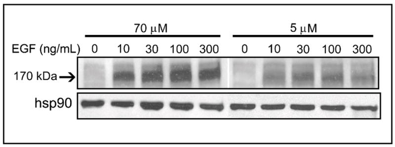
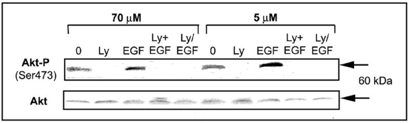
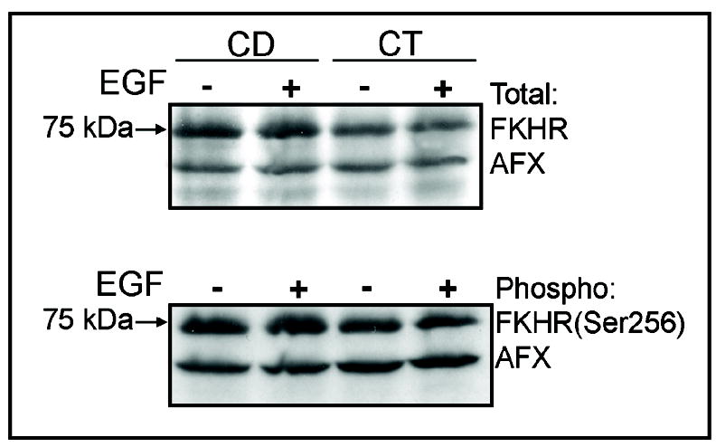
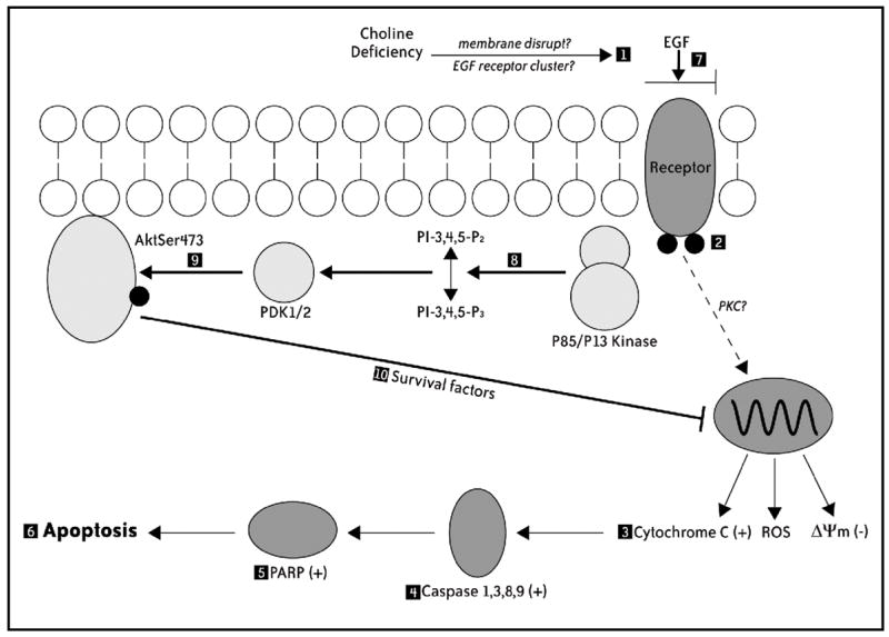
Previous studies show that acute choline deficiency (CD) triggers apoptosis in cultured rat hepatocytes (CWSV-1 cells). We demonstrate that prolonged EGF stimulation (10 ng/mL x 48 hrs) restores cell proliferation, as assessed by BrdU labeling, and protects cells from CD-induced apoptosis, as assessed by TUNEL labeling and cleavage of poly(ADP-ribose) polymerase. However, EGF rescue was not accompanied by restoration of depleted intracellular concentrations of choline, glycerphosphocholine, phosphocholine, or phosphatidylcholine. In contrast, we show that EGF stimulation blocks apoptosis by restoring mitochondrial membrane potential (Delta Psi(m)), as determined using the potential-sensitive dye chloromethyl-X-rosamine, and by preventing the release and nuclear localization of cytochrome c. We investigated whether EGF rescue involves EGF receptor phosphorylation and activation of the down-stream cell survival factor Akt. Compared to cells in control medium (CT, 70 micromol choline x 48 hrs), cells in CD medium (5 micromol choline) were less sensitive to EGF-induced (0-300 ng/mL x 5 min) receptor tyrosine phosphorylation. Compared to cells in CT medium, cells in CD medium treated with EGF (10 ng/mL x 5 min) exhibited higher levels of phosphatidylinositol 3-kinase (PI3K)-dependent phosphorylation of AktSer473. Inactivation of PI3K was sufficient to block EGF-stimulated activation of Akt, restoration of mitochondrial Delta Psi(m), and prevention of cytochrome c release. These studies indicate that stimulation with EGF activates a cell survival response against CD-apoptosis by restoring mitochondrial membrane potential and preventing cytochrome c release and nuclear translocation which are mediated by activation of Akt in hepatocytes.


Figures

Fig. 1
Fig. 1
Epidermal growth factor rescues p53-defective CWSV-1 cells from apoptosis induced by acute withdrawal of choline. CWSV-1 hepatocytes in the log phase of growth were switched for 2 days to RPMI medium containing 70 μmol choline (CT) or 5 μmol choline (CD) with or without epidermal growth factor (EGF) as described in the Materials and Methods. The occurrence of apoptosis was determined by (A) PARP cleavage and (B) a TUNEL assay for apoptosis-associated DNA fragmentation. Mean ± SE, n = 6/point; bars identified with different letters are significantly different from one another (p < 0.01, t-test).
Fig. 2
Fig. 2
Effects of choline availability and EGF on mitochondrial function. Mitochondrial transmembrane potential (ΔΨm) was studied as described in the Materials and Methods with the potential-sensitive probe CMX-Ros (red fluorescence) and the nuclear counterstain DAPI (blue fluorescence). Compared to CWSV-1 cells maintained in control medium (70 μmol choline) (A) cells maintained in choline deficient medium (5 μmol choline) exhibited decreased incorporation of CMX-Ros consistent with disruption of ΔΨm (C). Treatment with EGF (10ng/mL) for 48 hr restored MPT in CWSV-1 cells maintained in control (B) and choline deficient medium (D). The relative levels of CMX-Ros (E) were measured using an objective image analysis method as described in the Materials and Methods. In the absence of EGF, cells maintained in control medium (white bars) exhibited a 3-fold increase in the amount of CMX-Ros incorporated compared to cells in choline deficient medium (black bars). Treatment with EGF (10 ng/mL x 48hr) restored the level of incorporation of CMX-Ros in choline deficient cells to control levels. Consistent with restoration of ΔΨm. Mean ± SE, n = 4/treatment group; columns identified with different letters differ significantly from one another (p < 0.01, t-test).
Fig. 3
Fig. 3
Effects of choline availability and epidermal growth factor on cytochrome c release. CWSV-1 cells were grown and treated as described in Materials and Methods. Cells were fixed in formaldehyde and immunocytochemically stained for cytochrome c (red). The cells were visualized on a Nikon FX microscope and images captured by a digital camera. (A, B) Localization of cytochrome c staining in CT cells (70 μmol choline) grown in the absence (A) or presence (B) of EGF (10 ng/mL x 48 hrs). (C, D) Localization of cytochrome c staining in CD cells grown in the absence (C) or presence (D) of EGF. In the absence of EGF, CD cells exhibited a diffuse and cytoplasmic and nuclear pattern of cytochrome c staining, unlike the punctate perinuclear staining of cytochrome c in CT cells. Treatment with EGF (10 ng/mL x 48 hr) restored the punctate perinuclear pattern of cytochrome c staining in CD cells maintained in choline deficient medium (D) consistent with the pattern of staining observed in CT cells (B). (E) Cytochrome c levels in total cell lysates were unaffected by choline availability or EGF. Arrows: location of cytoplasm overlying nucleus.
Fig. 4
Fig. 4
Epidermal growth factor restores cell proliferation in CD-CWSV-1 cells. Cells in the log phase of growth were switched for an additional 2 days to control (70 μmol choline) or CD medium (5 μmol choline) with or without EGF, and the percentage of cells incorporating BrdU (BrdU labeling index) was determined as described in the Materials and Methods. Mean ± SE; columns identified with different letters differ significantly from one another (p < 0.01, ANOVA/Sheffe’s test).
Fig. 5
Fig. 5
Effects of choline availability on EGF receptor tyrosine phosphorylation. CWSV-1 cells were treated with EGF (0–300 ng/mL x 5 min) and the levels of tyrosine phosphorylation of EGF (EGF pTyr) was assayed immunoblotting with anti-phosphotyrosine antibody (RC-20) and quantified by densitometry, as described in the Materials and Methods. Hsp90 was used to normalize band intensities for protein loading. The arrow indicates the EGF pTyr band at ~ 170 kDa.
Fig. 6
Fig. 6
Effects of choline availability and activation of PI3 kinase on the expression of Akt. CWSV-1 cells were treated with EGF (10 ng/mL x 5 min) in the absence or presence of LY294002 and the levels of phosphorylated Akt and total Akt were assayed by immunoblotting with anti-Akt473 and total Akt antibodies, as described in the Materials and Methods. Treatment with EGF increased AktSer473 in CD medium (5 μM choline) cells more than in cells maintained in control medium (70 μM choline). Treatment with the LY294002 (50 μM) either simultaneous with (Ly+EGF) or for 1 hr before the addition of EGF (Ly/EGF) suppressed the expression of Akt, consistent with PI3 kinase-dependent activation of AktSer473 in response to EGF.
Fig. 7
Fig. 7
Effects of choline availability and EGF on the expression of Forkhead-related transcription factors. CWSV-1 cells were treated with EGF (10 ng/mL x 5 min) and the levels of phosphorylated and total FKHR and AFX were assayed by immunoblotting with antibodies that recognize FKHRSer256, total FKHR, AFXSer193, and total AFX., as described in the Materials and Methods. Cells in control (CT) and choline deficient medium (CD) exhibited similar levels of FKHR and AFX and treatment with EGF had no effect on the expression of phosphorylated (inactivated) or total Forkhead-related proteins.
Fig. 8
Fig. 8
Model for EGF-induced rescue against CD apoptosis in rat hepatocytes. Cells switched acutely to CD medium (1) exhibit decreased sensitivity to EGF-induced receptor tyrosine phosphorylation (2), and (3) proapoptotic mitochondrial alterations (i.e., loss of ΔΨm, cytochrome c release, increased generation of ROS) that result in (4) activation of a caspases cascade, (5) cleavage of PARP, and (6) the completion of apoptosis. In the presence of added EGF (7), CD cells exhibit (8) PI3 kinase-dependent activation of AktSer473 (9), and (10) restoration of early mitochondrial pro-apoptotic alterations (i.e., ΔΨm, cytochrome c), independent of the expression of Forkhead-related proteins (i.e., FKHR, AFX) and phosphorylated Bad.

References

    1. Food and Nutrition Board, Institute of Medicine. Dietary References Intakes for Thiamin, Riboflavin, Niacin, Vitamin B6, Folate, Vitamin B12, Pantothenic Acid, Biotin and Choline. National Academy Press; Washington, D.C: 1998. - PubMed
    1. Blusztajn JK. Choline: a vital amine. Science. 1998;281:794–795. - PubMed
    1. Jackowski S. Coordination of membrane phospholipid synthesis with the cell cycle. J Biol Chem. 1994;269:3858–3867. - PubMed
    1. Zeisel SH. Choline: essential for brain development and function. Adv Pediatr. 1997;44:263–295. - PubMed
    1. Albright CD, Liu R, Bethea TC, da Costa K-A, Salganik RI, Zeisel SH. Choline deficiency induces apoptosis in CWSV-1 hepatocytes in culture. FASEB J. 1996;10:510–516. - PubMed

Publication types

MeSH terms