

A comparative study on the acute and long-term effects of MDMA and 3,4-dihydroxymethamphetamine (HHMA) on brain monoamine levels after i.p. or striatal administration in mice
- PMID: 15665862
- PMCID: PMC1575997
- DOI: 10.1038/sj.bjp.0706071
A comparative study on the acute and long-term effects of MDMA and 3,4-dihydroxymethamphetamine (HHMA) on brain monoamine levels after i.p. or striatal administration in mice
Abstract
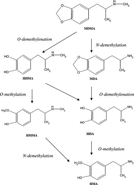
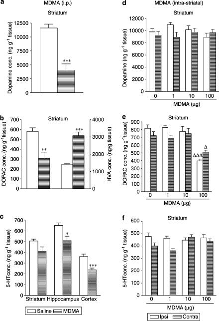
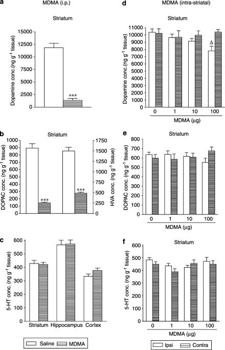
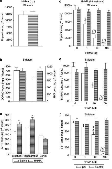
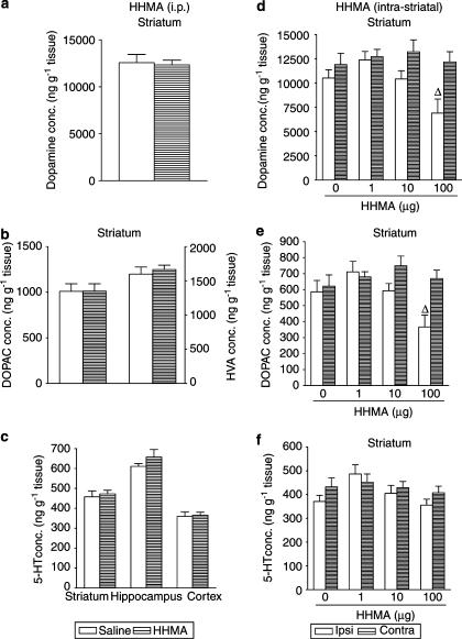
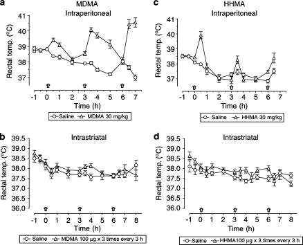
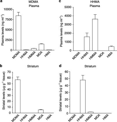
1. This study investigated whether the immediate and long-term effects of 3,4-methylenedioxymethamphetamine (MDMA) on monoamines in mouse brain are due to the parent compound and the possible contribution of a major reactive metabolite, 3,4-dihydroxymethamphetamine (HHMA), to these changes. The acute effect of each compound on rectal temperature was also determined. 2. MDMA given i.p. (30 mg kg(-1), three times at 3-h intervals), but not into the striatum (1, 10 and 100 microg, three times at 3-h intervals), produced a reduction in striatal dopamine content and modest 5-HT reduction 1 h after the last dose. MDMA does not therefore appear to be responsible for the acute monoamine release that follows its peripheral injection. 3. HHMA does not contribute to the acute MDMA-induced dopamine depletion as the acute central effects of MDMA and HHMA differed following i.p. injection. Both compounds induced hyperthermia, confirming that the acute dopamine depletion is not responsible for the temperature changes. 4. Peripheral administration of MDMA produced dopamine depletion 7 days later. Intrastriatal MDMA administration only produced a long-term loss of dopamine at much higher concentrations than those reached after the i.p. dose and therefore bears little relevance to the neurotoxicity. This indicates that the long-term effect is not attributable to the parent compound. HHMA also appeared not to be responsible as i.p. administration failed to alter the striatal dopamine concentration 7 days later. 5. HHMA was detected in plasma, but not in brain, following MDMA (i.p.), but it can cross the blood-brain barrier as it was detected in the brain following its peripheral injection. 6. The fact that the acute changes induced by i.p. or intrastriatal HHMA administration differed indicates that HHMA is metabolised to other compounds which are responsible for changes observed after i.p. administration.
Figures
Similar articles
-
Effects of intracerebroventricular administration of 5-(glutathion-S-yl)-alpha-methyldopamine on brain dopamine, serotonin, and norepinephrine concentrations in male Sprague-Dawley rats.Chem Res Toxicol. 1996 Mar;9(2):457-65. doi: 10.1021/tx9501546. Chem Res Toxicol. 1996. PMID: 8839050
-
Effect of acute brain tyrosine depletion on MDMA-induced changes in brain 5-HT.J Psychopharmacol. 2010 Feb;24(2):267-74. doi: 10.1177/0269881109348163. Epub 2009 Dec 4. J Psychopharmacol. 2010. PMID: 19965941
-
A study of the mechanisms involved in the neurotoxic action of 3,4-methylenedioxymethamphetamine (MDMA, 'ecstasy') on dopamine neurones in mouse brain.Br J Pharmacol. 2001 Dec;134(8):1711-23. doi: 10.1038/sj.bjp.0704435. Br J Pharmacol. 2001. PMID: 11739248 Free PMC article.
-
Reinforcing effects of methylenedioxy amphetamine congeners in rhesus monkeys: are intravenous self-administration experiments relevant to MDMA neurotoxicity?Psychopharmacology (Berl). 2007 Jan;189(4):471-82. doi: 10.1007/s00213-006-0320-8. Epub 2006 Mar 23. Psychopharmacology (Berl). 2007. PMID: 16555062 Review.
-
The preclinical pharmacology of mephedrone; not just MDMA by another name.Br J Pharmacol. 2014 May;171(9):2251-68. doi: 10.1111/bph.12628. Br J Pharmacol. 2014. PMID: 24654568 Free PMC article. Review.
Cited by
-
Role of the dopaminergic system in the acquisition, expression and reinstatement of MDMA-induced conditioned place preference in adolescent mice.PLoS One. 2012;7(8):e43107. doi: 10.1371/journal.pone.0043107. Epub 2012 Aug 16. PLoS One. 2012. PMID: 22916213 Free PMC article.
-
Neuronal and peripheral damages induced by synthetic psychoactive substances: an update of recent findings from human and animal studies.Neural Regen Res. 2020 May;15(5):802-816. doi: 10.4103/1673-5374.268895. Neural Regen Res. 2020. PMID: 31719240 Free PMC article. Review.
-
Catechol-o-methyltransferase and 3,4-({+/-})-methylenedioxymethamphetamine toxicity.Toxicol Sci. 2014 May;139(1):162-73. doi: 10.1093/toxsci/kfu035. Epub 2014 Mar 3. Toxicol Sci. 2014. PMID: 24591155 Free PMC article.
-
Administration of neurotoxic doses of MDMA reduces sensitivity to ethanol and increases GAT-1 immunoreactivity in mice striatum.Psychopharmacology (Berl). 2010 Jan;207(4):671-9. doi: 10.1007/s00213-009-1699-9. Epub 2009 Oct 20. Psychopharmacology (Berl). 2010. PMID: 19841904
-
Cocaine potentiates MDMA-induced oxidative stress but not dopaminergic neurotoxicity in mice: implications for the pathogenesis of free radical-induced neurodegenerative disorders.Psychopharmacology (Berl). 2013 Nov;230(1):125-35. doi: 10.1007/s00213-013-3142-5. Epub 2013 May 17. Psychopharmacology (Berl). 2013. PMID: 23681166
References
-
- BUCHERT R., THOMASIUS R., NEBELING B., PETERSEN K., OBROCKI J., JENICKE L., WILKE F., WARTBERG L., ZAPLETALOVA P., CLAUSEN M. Long-term effects of ‘ecstasy' use on serotonin transporters of the brain investigated by PET. J. Nucl. Med. 2003;44:375–384. - PubMed
-
- CAMARERO J., SANCHEZ V., O'SHEA E., GREEN A.R., COLADO M.I. Studies, using in vivo microdialysis, on the effect of the dopamine uptake inhibitor GBR 12909 on 3,4-methylenedioxymethamphetamine (‘ecstasy')-induced dopamine release and free radical formation in the mouse striatum. J. Neurochem. 2002;81:961–972. - PubMed
-
- CHO A.K., HIRAMATSU M., DISTEFANO E.W., CHANG A.S., JENDEN D.J. Stereochemical differences in the metabolism of 3,4-methylenedioxymethamphetamine in vitro and in vivo: a pharmacokinetic analysis. Drug Metab. Disposition. 1990;18:686–691. - PubMed
Publication types
MeSH terms
Substances
LinkOut - more resources
Full Text Sources
Medical
