

doi: 10.1002/anie.200462114.
Recognizing a single base in an individual DNA strand: a step toward DNA sequencing in nanopores
Affiliations
- PMID: 15666419
- PMCID: PMC1828035
- DOI: 10.1002/anie.200462114
Item in Clipboard
Recognizing a single base in an individual DNA strand: a step toward DNA sequencing in nanopores
Angew Chem Int Ed Engl.
.
Abstract
Functional supramolecular chemistry at the single-molecule level. Single strands of DNA can be captured inside α-hemolysin transmembrane pore protein to form single-species α-HL·DNA pseudorotaxanes. This process can be used to identify a single adenine nucleotide at a specific location on a strand of DNA by the characteristic reductions in the α-HL ion conductance. This study suggests that α-HL-mediated single-molecule DNA sequencing might be fundamentally feasible.
Figures
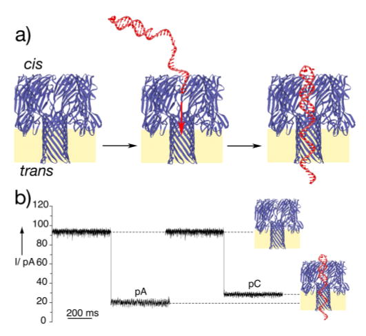
a) Schematic representation of the single-species α–
HL·DNA pseudorotaxane formation. By employing sequences with a
long ss-DNA segment having a hairpin structure at one end, the SS-DNA can
thread the pore under positive applied potentials and held stably in a
pseudorotaxane configuration by the interactions of the DNA duplex segment
at the pore entrance. The heptameric transmembrane pore structure is
depicted in a cutaway side view. b) Ion conductance blockades caused by
capture of poly d(A) and poly d(C) single strands (sequences 1
and 2, respectively). The typical events shown are from ion
conductance traces recorded at +170 mV when the thread molecules
(1 μM) were added to the cis chamber. Traces
were recorded under symmetrical conditions (500 mM KCl, 5 mM MOPS pH 7.5),
Bessel filtered at 5 kHz, and sampled at 200 μs.
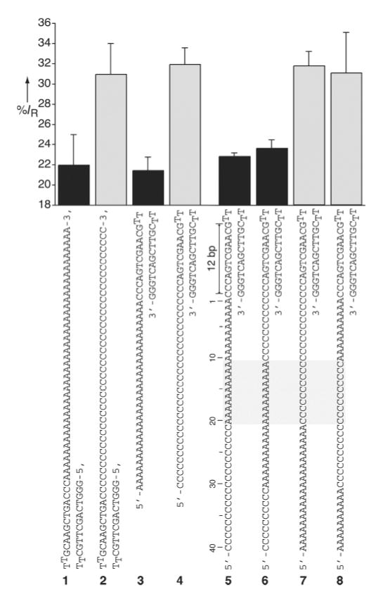
The averaged residual currents for various α-HL·DNA
pseudorotaxanes measured at +170 mV. The left panel depicts the
percent residual currents measured for poly d(A) and poly d(C) threads in
two topoisomeric α-HL·DNA pseudorotaxane
configurations. Note that when the α-HL pore is threaded with a
homopolymeric DNA segment either from the free 3’-terminus
(1 or 2) or from 5’-terminus
(3 or 4), similar characteristic A-type or
C-type ion channel blockades are observed. The right panel depicts the
observed residual currents for α-HL·DNA pseudorotaxanes
prepared using four different ss-DNA block copolymer threads
(5–6). The region of the ss-DNA segment
recognized by the α-HL channel (10 to 20 nucleotides away from the
edge of the hairpin segment) is marked by the gray rectangle. For clarity
A-type and C-type residual currents are shown as black and gray bars,
respectively.
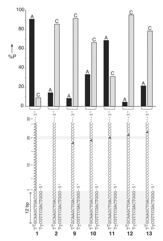
Percent probability of the A-type (IR < 27%)
and the C-type (IR > 27%) residual currents
for single deoxyadenosine-substituted poly d(C) DNA strands captured inside
the α-HL pore as a pseudorotaxane. Percent probability was
calculated by the number of A-type or C-type events measured at
+170 mV divided by the total number of events (n) recorded for a
given strand (n = 11, 21, 22, 32, 22, and 14 for strands
1, 2, 9, 10, 11, 12,
13, respectively). The percent probability of A-type and
C-type events for each strand are given as black and gray bars
respectively.
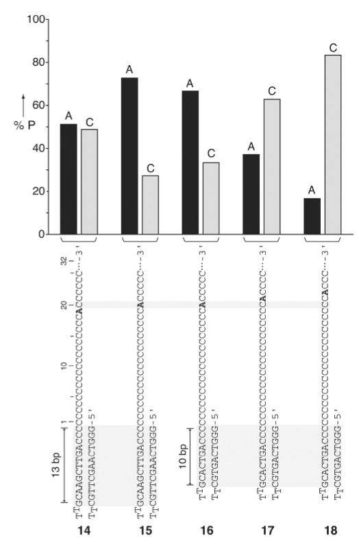
Percent probability of the A-type and the C-type events for single
deoxyadenosine-substituted poly d(C) DNA hairpins sequences having either
13- or 10-basepair stem duplexes (strands
14–15, and
16–18, respectively) measured at +170
mV (n = 43, 44, 69, 35, 66 for
14–18, respectively). For clarity
the A-type (IR < 27%) and C-type
(IR > 27%) events are shown as black and grey
bars, respectively.
References
-
- Song L, Hobaugh MR, Shustak C, Cheley S, Bayley H, Gouaux JE. Science. 1996;274:1859. - PubMed
-
- Kasianowicz JJ, Brandin E, Branton D, Deamer DW. Proc Natl Acad Sci USA. 1996;93:13770. - PMC - PubMed
- Akeson M, Branton D, Kasianowicz JJ, Brandin E, Deamer DW. Biophys J. 1999;77:3227. - PMC - PubMed
- Meller A, Nivon L, Brandin E, Golovchenko J, Branton D. Proc Natl Acad Sci USA. 2000;97:1079. - PMC - PubMed
- Howorka S, Cheley S, Bayley H. Nat Biotechnol. 2001;19:636. - PubMed
-
- Jirage KB, Hulteen JC, Martin CR. Science. 1997;278:655.
- Schmidt C, Mayer M, Vogel H. Angew Chem. 2000;112:3267. Angew. Chem. Int. Ed. 2000, 39, 3137. - PubMed
- Li J, Stein D, McMullan C, Branton D, Aziz MJ, Golovchenko JA. Nature. 2001;412:166. - PubMed
- Bayley H, Cremer PS. Nature. 2001;413:226. - PubMed
- Chen P, Mitsui T, Farmer DB, Golovchenko J, Gordon RG, Branton D. Nano Lett. 2004;4:1333. - PMC - PubMed
- Chang H, Kosari F, Andreadakis G, Alam MA, Vasmatzis G, Bashir R. Nano Lett. 2004;4:1551.
-
- Sauer-Budge AF, Nyamwanda JA, Lubensky DK, Branton D. Phys Rev Lett. 2003;90:238101. - PubMed
- Sanchez-Quesada J, Saghatelian A, Cheley S, Bayley H, Ghadiri MR. Angew Chem Int Ed. 2004;43:3063. Angew. Chem. 2004, 116, 3125. - PMC - PubMed
- Nakane J, Wiggin M, Marziali A. Biophys J. 2004;87:615. - PMC - PubMed
Publication types
MeSH terms
Substances
Grants and funding
LinkOut - more resources
Full Text Sources
Other Literature Sources
