

Antibody repertoire development in fetal and neonatal piglets. XI. The thymic B-cell repertoire develops independently from that in blood and mesenteric lymph nodes
- PMID: 15667562
- PMCID: PMC1782081
- DOI: 10.1111/j.1365-2567.2004.02101.x
Antibody repertoire development in fetal and neonatal piglets. XI. The thymic B-cell repertoire develops independently from that in blood and mesenteric lymph nodes
Abstract
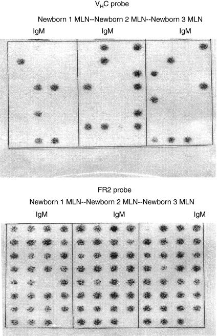
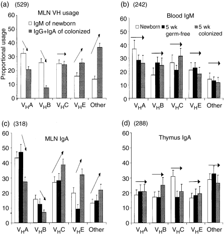
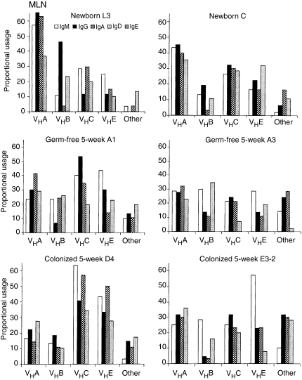
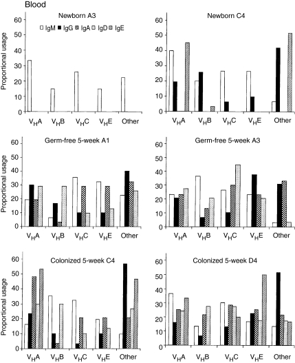
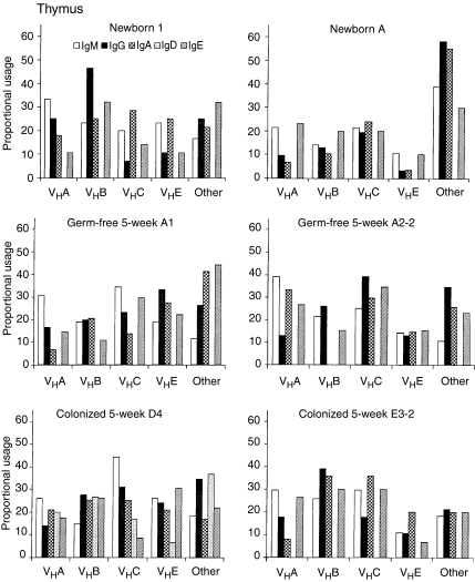
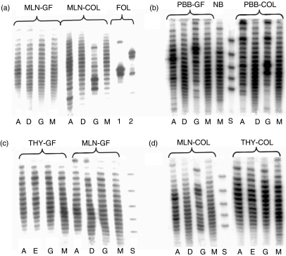
The origin and function of thymic B cells is currently unresolved. In the present study we compared V(H) gene repertoire diversification in >3500 cloned VDJs (from 11 animals at three time-points, using three to five animals per time-point) that were expressed with immunoglobulin (Ig)M, IgD, IgG, IgA and IgE in thymus, mesenteric lymph nodes (MLN) and peripheral blood B cells (PBB) of newborn piglets and 5-week-old isolator piglets maintained germfree (GF) or colonized with Escherichia coli. The results showed that the repertoire expressed with IgM, IgD, IgG and IgA in PBB and MLN remained polyclonal, undiversified and unselected in piglets maintained GF for 5 weeks, that age and colonization resulted in significant repertoire diversification of IgG and IgA in the MLN and of IgG in blood, that the thymic B-cell repertoire was polyclonal, unaffected by colonization and showed no clonal selection in any isotype, and that the thymic IgA and IgE repertoires were more diverse at birth than the repertoire of any isotype in MLN or PBB. IgD was seldom recovered from the PBB of newborn piglets or at any time-point in thymus, but was recovered in the MLN of all 11 animals examined. The IgD and IgM repertoires in all tissues remained polyclonal and unselected, although V(H) usage by IgD transcripts did not always parallel that of IgM in the same tissue. Therefore, isotype-switched B cells in the thymic medulla cannot be accounted for by immigration of B cells diversified by colonization of the gut, and thymic B cells undergo switch recombination and repertoire diversification before birth without clonal selection.
Figures
Similar articles
-
Antibody repertoire development in fetal and neonatal piglets. IV. Switch recombination, primarily in fetal thymus, occurs independent of environmental antigen and is only weakly associated with repertoire diversification.J Immunol. 2001 Sep 15;167(6):3239-49. doi: 10.4049/jimmunol.167.6.3239. J Immunol. 2001. PMID: 11544311
-
Antibody repertoire development in fetal and newborn piglets, III. Colonization of the gastrointestinal tract selectively diversifies the preimmune repertoire in mucosal lymphoid tissues.Immunology. 2000 May;100(1):119-30. doi: 10.1046/j.1365-2567.2000.00013.x. Immunology. 2000. PMID: 10809967 Free PMC article.
-
Thymic B cells of pig fetuses and germ-free pigs spontaneously produce IgM, IgG and IgA: detection by ELISPOT method.Immunology. 1996 Mar;87(3):487-92. doi: 10.1046/j.1365-2567.1996.499573.x. Immunology. 1996. PMID: 8778038 Free PMC article.
-
Antibody repertoire development in swine.Dev Comp Immunol. 2006;30(1-2):199-221. doi: 10.1016/j.dci.2005.06.025. Dev Comp Immunol. 2006. PMID: 16168480 Review.
-
Antibody Repertoire Development in Swine.Annu Rev Anim Biosci. 2017 Feb 8;5:255-279. doi: 10.1146/annurev-animal-022516-022818. Annu Rev Anim Biosci. 2017. PMID: 28199170 Review.
Cited by
-
Porcine IgG: structure, genetics, and evolution.Immunogenetics. 2009 Mar;61(3):209-30. doi: 10.1007/s00251-008-0336-9. Epub 2008 Dec 2. Immunogenetics. 2009. PMID: 19048248
-
The piglet as a model for B cell and immune system development.Vet Immunol Immunopathol. 2009 Mar 15;128(1-3):147-70. doi: 10.1016/j.vetimm.2008.10.321. Epub 2008 Nov 1. Vet Immunol Immunopathol. 2009. PMID: 19056129 Free PMC article. Review.
-
The isolator piglet: a model for studying the development of adaptive immunity.Immunol Res. 2007;39(1-3):33-51. doi: 10.1007/s12026-007-0062-7. Immunol Res. 2007. PMID: 17917054 Review.
-
The ontogeny of the porcine immune system.Dev Comp Immunol. 2009 Mar;33(3):273-83. doi: 10.1016/j.dci.2008.07.011. Epub 2008 Aug 30. Dev Comp Immunol. 2009. PMID: 18762210 Free PMC article. Review.
-
Thymic B cell development is controlled by the B potential of progenitors via both hematopoietic-intrinsic and thymic microenvironment-intrinsic regulatory mechanisms.PLoS One. 2018 Feb 20;13(2):e0193189. doi: 10.1371/journal.pone.0193189. eCollection 2018. PLoS One. 2018. PMID: 29462202 Free PMC article.
References
-
- Sainte-Marie G. Plasmacytes in the thymus of the normal rat. J Immunol. 1965;94:172–4. - PubMed
-
- Butler JE, Maxwell CF, Pierce CS, Hylton MB, Asofsky R, Kiddy CA. Studies on the relative synthesis and distribution of IgA and IgG1 in various tissues and body fluids of the cow. J Immunol. 1972;109:38–46. - PubMed
-
- Dixon FJ, Weigle WO, Roberts JC. Comparison of antibody responses associated with the transfer of rabbit lymph node, peritoneal exudate and thymus cells. J Immunol. 1957;78:56–63. - PubMed
-
- Butler JE, Sun J, Weber P, Ford SP, Rehakova Z, Sinkora J, Lager K. Antibody repertoire development in fetal and neonatal piglets. IV. Switch recombination, primary in fetal thymus occurs independent of environmental antigen and is only weakly associated with repertoire diversification. J Immunol. 2001;167:3239–49. - PubMed
Publication types
MeSH terms
Substances
LinkOut - more resources
Full Text Sources
Miscellaneous
