

Mucosal delivery of CpG oligodeoxynucleotides expands functional dendritic cells and macrophages in the vagina
- PMID: 15667566
- PMCID: PMC1782077
- DOI: 10.1111/j.1365-2567.2004.02081.x
Mucosal delivery of CpG oligodeoxynucleotides expands functional dendritic cells and macrophages in the vagina
Abstract
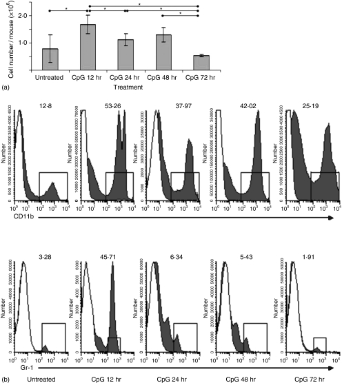
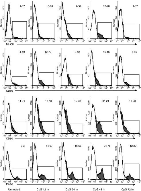
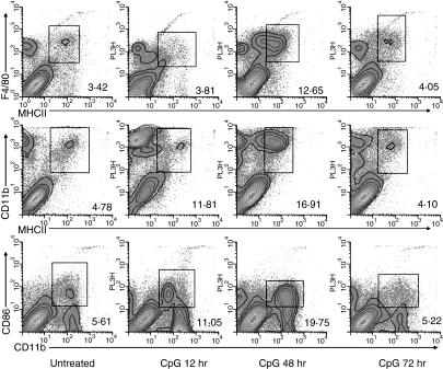
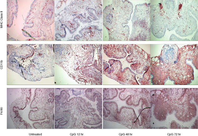
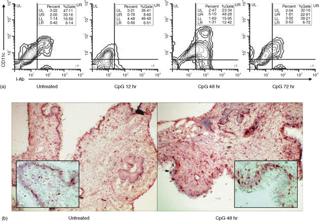
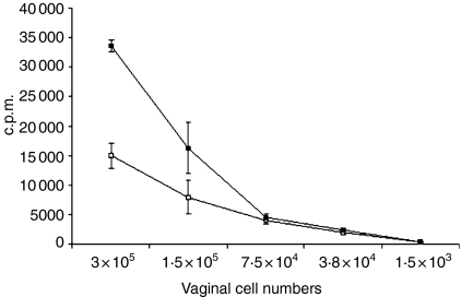
Antigen-presenting cells (APC) are specialized sentinel cells that sense pathogens within tissues and then activate appropriate immune effector cells in lymphoid organs. Recent evidence, however, suggests that APC can also induce effector cells in non-lymphoid organs. The purpose of this study was to determine the effect of intravaginal (IVAG) delivery of CpG-oligodeoxynucleotide (ODN) on expansion of resident genital APC. Our results show that delivery of CpG-ODN to the murine genital tract induced a rapid and significant, but transient expansion of genital APC in situ. As early as 12 hr post CpG-ODN delivery, we observed an enhanced level of F4/80+ major histocompatibility complex (MHC) class II-negative macrophages in the genital tissue. This was followed by increased levels of F4/80/MHC class II double-positive cells, as well as MHC class II, CD11c and CD86 triple-positive dendritic cells (DC) at 48 hr. Expanded APC levels at 48 hr post CpG-ODN resulted in increased ability of genital cells to induce an allogenic mixed leucocyte reaction. By 72 hr after CpG-ODN treatment, APC levels were not distinguishable from naive levels. Therefore, these results clearly show that administration of CpG-ODN to the genital tract induced a marked but transient enhancement of APC within the genital tissue, and that these APC appear to possess functional capacity. Furthermore, these results indicate that IVAG-CpG-ODN may be an important factor for the enhancement of local antigen presentation in the genital tract through increased DC numbers.
Figures
Similar articles
-
Intravaginal immunization with viral subunit protein plus CpG oligodeoxynucleotides induces protective immunity against HSV-2.Vaccine. 2004 Aug 13;22(23-24):3098-104. doi: 10.1016/j.vaccine.2004.01.059. Vaccine. 2004. PMID: 15297061
-
Parameters of CpG oligodeoxynucleotide-induced protection against intravaginal HSV-2 challenge.J Med Virol. 2003 Dec;71(4):561-8. doi: 10.1002/jmv.10518. J Med Virol. 2003. PMID: 14556270
-
CpG oligodeoxynucleotides down-regulate macrophage class II MHC antigen processing.J Immunol. 1999 Aug 1;163(3):1188-94. J Immunol. 1999. PMID: 10415013
-
CD8+ T-cell-mediated cross-clade protection in the genital tract following intranasal immunization with inactivated human immunodeficiency virus antigen plus CpG oligodeoxynucleotides.J Virol. 2005 Jan;79(1):393-400. doi: 10.1128/JVI.79.1.393-400.2005. J Virol. 2005. PMID: 15596832 Free PMC article.
-
Bacterial antigen delivery systems: phagocytic processing of bacterial antigens for MHC-I and MHC-II presentation to T cells.Behring Inst Mitt. 1997 Feb;(98):197-211. Behring Inst Mitt. 1997. PMID: 9382741 Review.
Cited by
-
A review of HSV pathogenesis, vaccine development, and advanced applications.Mol Biomed. 2024 Aug 29;5(1):35. doi: 10.1186/s43556-024-00199-7. Mol Biomed. 2024. PMID: 39207577 Free PMC article. Review.
-
Effect of Mucosal Cytokine Administration on Selective Expansion of Vaginal Dendritic Cells to Support Nanoparticle Transport.Am J Reprod Immunol. 2015 Oct;74(4):333-44. doi: 10.1111/aji.12409. Epub 2015 Jun 27. Am J Reprod Immunol. 2015. PMID: 26118309 Free PMC article.
-
Inhibitory effect of PRO 2000, a candidate microbicide, on dendritic cell-mediated human immunodeficiency virus transfer.Antimicrob Agents Chemother. 2008 May;52(5):1751-8. doi: 10.1128/AAC.00707-07. Epub 2008 Mar 10. Antimicrob Agents Chemother. 2008. PMID: 18332174 Free PMC article.
-
Engineering immunity in the mucosal niche against sexually transmitted infections.Wiley Interdiscip Rev Nanomed Nanobiotechnol. 2016 Jan-Feb;8(1):107-22. doi: 10.1002/wnan.1359. Epub 2015 Jul 7. Wiley Interdiscip Rev Nanomed Nanobiotechnol. 2016. PMID: 26153141 Free PMC article. Review.
-
Unraveling molecular signatures of immunostimulatory adjuvants in the female genital tract through systems biology.PLoS One. 2011;6(6):e20448. doi: 10.1371/journal.pone.0020448. Epub 2011 Jun 7. PLoS One. 2011. PMID: 21666746 Free PMC article.
References
-
- Steinman RM. The dendritic cell system and its role in immunogenicity. Annu Rev Immunol. 1991;9:271. 10.1146/annurev.iy.09.040191.001415. - DOI - PubMed
-
- Fernandez NC, Lozier A, Flament C, et al. Dendritic cells directly trigger NK cell functions: cross-talk relevant in innate anti-tumor immune responses in vivo. Nat Med. 1999;5:405. 10.1038/7403. - DOI - PubMed
-
- Bienenstock J, Johnston N, Perey DY. Bronchial lymphoid tissue. I. Morphologic characteristics. Lab Invest. 1973;28:686. - PubMed
-
- Bienenstock J, Johnston N, Perey DY. Bronchial lymphoid tissue. II. Functional characterisitics. Lab Invest. 1973;28:693. - PubMed
Publication types
MeSH terms
Substances
LinkOut - more resources
Full Text Sources
Research Materials
