. 2005 Feb;11(2):245-56.
doi: 10.1016/j.ymthe.2004.09.013.

Adeno-associated virus-mediated microdystrophin expression protects young mdx muscle from contraction-induced injury

Affiliations

Adeno-associated virus-mediated microdystrophin expression protects young mdx muscle from contraction-induced injury

Mingju Liu et al. Mol Ther. 2005 Feb.

Abstract

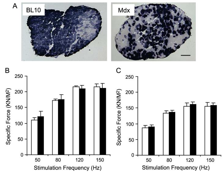
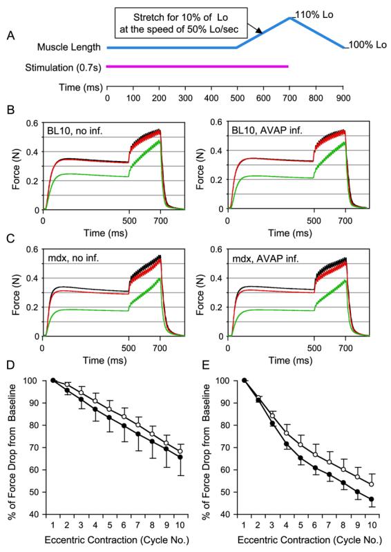
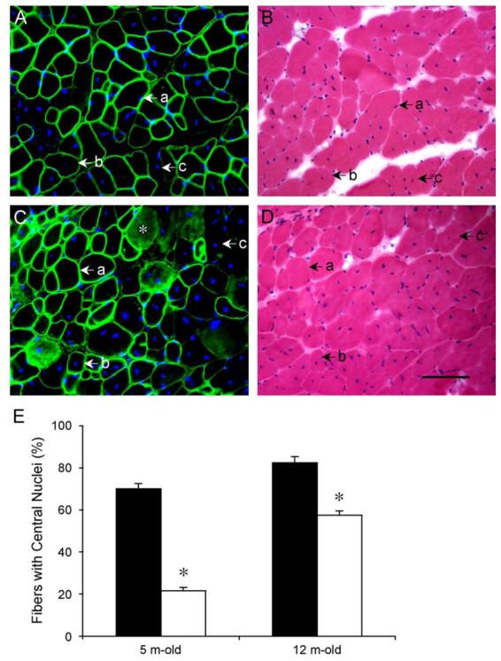
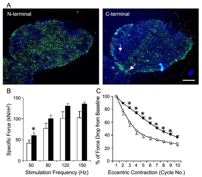
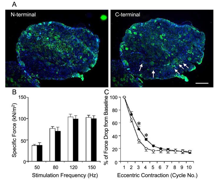
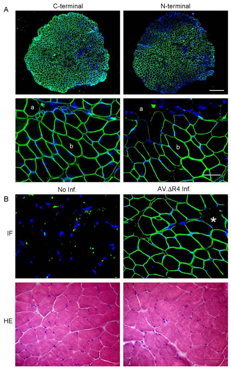
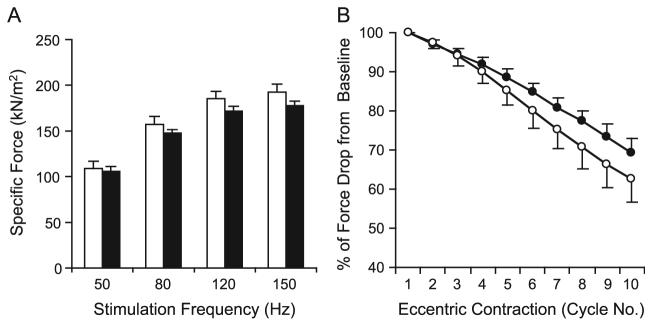
Duchenne muscular dystrophy (DMD) is the most common inherited lethal muscle degenerative disease. Currently there is no cure. Highly abbreviated microdystrophin cDNAs were developed recently for adeno-associated virus (AAV)-mediated DMD gene therapy. Among these, a C-terminal-truncated DeltaR4-R23/DeltaC microgene (DeltaR4/DeltaC) has been considered as a very promising therapeutic candidate gene. In this study, we packaged a CMV.DeltaR4/DeltaC cassette in AAV-5 and evaluated the transduction and muscle contractile profiles in the extensor digitorum longus muscles of young (7-week-old) and adult (9-month-old) mdx mice. At approximately 3 months post-gene transfer, 50-60% of the total myofibers were transduced in young mdx muscle and the percentage of centrally nucleated myofibers was reduced from approximately 70% in untreated mdx muscle to approximately 22% in microdystrophin-treated muscle. Importantly, this level of transduction protected mdx muscle from eccentric contraction-induced damage. In contrast, adult mdx muscle was more resistant to AAV-5 transduction, as only approximately 30% of the myofibers were transduced at 3 months postinfection. This transduction yielded marginal protection against eccentric contraction-induced injury. The extent of central nucleation was also more difficult to reverse in adult mdx muscle (from approximately 83% in untreated to approximately 58% in treated). Finally, we determined that the DeltaR4/DeltaC microdystrophin did not significantly alter the expression pattern of the endogenous full-length dystrophin in normal muscle. Neither did it have any adverse effects on normal muscle morphology or contractility. Taken together, our results suggest that AAV-mediated DeltaR4/DeltaC microdystrophin expression represents a promising approach to rescue muscular dystrophy in young mdx skeletal muscle.


Figures

FIG. 1
FIG. 1
AV.RSV.AP infection did not reduce isometric tetanic force in the EDL muscle. The left EDL muscles of 6-week-old mice were infected with AV.RSV.AP and the contralateral right EDL muscles were injected with equal volumes of saline. Transgene expression and tetanic force were evaluated at 4 weeks postinfection. (A) Efficient AAV transduction was observed in both normal (BL10) and dystrophic (mdx) EDL muscles. Scale bar, 200 μm. (B) Force–frequency relationship in BL10 EDL muscles. N = 4 pairs. (C) Force–frequency relationship in mdx EDL muscles. N = 5 pairs. Open bar, without AAV infection; filled bar, infected with AV.RSV.AP. AAV infection did not alter specific force production in BL10 and mdx EDL muscles.
FIG. 2
FIG. 2
AV.RSV.AP infection did not aggravate eccentric contraction-induced injury in the EDL muscle. (A) Schematic outline of the eccentric contraction protocol used in this study. Muscle was stimulated at 150 Hz for 700 ms (pink line). At the beginning of the stimulation, muscle length (blue line) was adjusted to the optimal length (Lo). At the end of 500 ms stimulation, muscle length was stretched to 110% of Lo at the speed of 50% Lo/s. At the end of stimulation, muscle length was returned to Lo at the same speed. A total of 10 stretch (eccentric contraction) cycles were performed in each muscle. An isometric tetanic force was developed during the first 500 ms stimulation. The change of this tetanic force between each eccentric contraction cycle reflected the degree of muscle injury. (B) Representative force tracing in BL10 EDL muscles. Left, without AAV infection; right, infected with AV.RSV.AP. Black line, force tracing from the first cycle. Red line, force tracing from the second cycle. Green line, force tracing from the 10th cycle. (C) Representative force tracing in mdx EDL muscles. Left, without AAV infection; right, infected with AV.RSV.AP. Black line, force tracing from the first cycle. Red line, force tracing from the second cycle. Green line, force tracing from the 10th cycle. The isometric tetanic force drop was more significant in mdx muscles. However, there was no significant difference between the AV.RSV.AP-infected muscle and the saline-injected control. (D) Relative change in tetanic force during 10 cycles of eccentric contraction in BL10 EDL muscles (N = 4 pairs). The tetanic tension developed during the first cycle was designated as 100%. Open circle, without AAV infection (mean + SEM). Closed circle, infected with AV.RSV.AP (mean − SEM). (E) Relative change of tetanic force during 10 cycles of eccentric contraction in mdx EDL muscles (N = 5 pairs). The isometric tetanic tension developed during the first cycle was designated as 100%. Open circle, without AAV infection (mean + SEM). Closed circle, infected with AV.RSV.AP (mean − SEM). AV.RSV.AP infection resulted in a slightly bigger, but not statistically significant, force deficit in the last few cycles. (For interpretation of the references to color in this figure legend, the reader is referred to the web version of this article.)
FIG. 3
FIG. 3
AAV-mediated microdystrophin expression was more efficient in reducing central nucleation in young (7-week-old) mdx EDL muscles. AV.ΔR4/ΔC was delivered to EDL muscles of 7-week-old (young) and 9-month-old (adult) mdx mice. Three months later, the percentage of centrally nucleated myofibers in all transduced (ΔR4/ΔC-positive) myofibers was quantified in each infected EDL muscle. (A and B) Representative immunofluorescence (A, with microdystrophin-specific antibody) and HE (B) staining of an EDL muscle infected at 7 weeks of age. (a) A microdystrophin-positive fiber with a peripherally located nucleus. (b) A microdystrophin-positive fiber with a centrally located nucleus. (c) A microdystrophin-negative fiber with a centrally located nucleus. (C and D) Representative immunofluorescence (C, with microdystrophin-specific antibody) and HE (D) staining of an EDL muscle infected at 9 months of age. (a) A microdystrophin-positive fiber with a peripherally located nucleus. (b) A microdystrophin-positive fiber with a centrally located nucleus. (c) A microdystrophin-negative fiber with a centrally located nucleus. Nonspecific immunoreactivity was observed in cytosol of some myofibers in old mdx muscle (*). In A and C, nuclei were stained with DAPI. Scale bar for D (100 μm) applies to all the photomicrographs. (E) Quantitative evaluation of central nucleation in young (infected at 7 weeks of age and examined at 5 months of age) and older (infected at 9 months of age and examined at 12 months of age) EDL muscles. Filled bar, percentage of fibers with centrally located nuclei in untreated mdx muscles. Open bar, percentage of fibers with centrally located nuclei in microdystrophin-positive myofibers in AV.ΔR4/ΔC-infected mdx muscles. N = 5 for all groups except for 5-month-old AV.ΔR4/ΔC-infected group (N = 9). There was a statistically significant difference between untreated and treated muscles (*P < 0.05). Muscles infected at the younger age were better protected than those infected at the older age.
FIG. 4
FIG. 4
AAV-mediated ΔR4/ΔC microdystrophin expression protected young (7 weeks old at infection) EDL muscles from eccentric contraction-induced injury. The left EDL muscles of 7-week-old mdx mice were infected with AV.ΔR4/ΔC. The contralateral right EDL muscles were infected with AV.RSV.AP. Viral transduction and muscle physiology were examined when mice were 5 months of age. (A) Representative photomicrographs of immunofluorescence staining with the N-terminal-specific (for ΔR4/ΔC microdystrophin, left) and the C-terminal-specific (for endogenous revertant murine dystrophin, right) antibodies. Nuclei were stained with DAPI. Scale bar, 300 μm. On average, approximately 58% of EDL myofibers were transduced by AAV (Table 2). Revertant myofibers were occasionally seen with the antibody against the dystrophin C-terminus (arrow, right). (B) Effect of partial microdystrophin transduction on specific tetanic force in the mdx EDL muscle. Open bar, EDL muscles infected with AV.RSV.AP; filled bar, EDL muscles infected with AV.ΔR4/ΔC. N = 5 pairs. *The difference between AV.ΔR4/ΔC- and AV.RSV.AP-infected muscles was statistically significant ( P < 0.05). (C) Partial microdystrophin expression protected the EDL muscle from the majority of eccentric contraction-induced injuries (from the third to the ninth cycle). Open circle, EDL muscles infected with AV.RSV.AP (mean − SEM); closed circle, EDL muscles infected with AV.ΔR4/ΔC (mean + SEM). N = 5 pairs. *The difference between AV.ΔR4/ΔC- and AV.RSV.AP-infected muscles was statistically significant ( P < 0.05).
FIG. 5
FIG. 5
Adult (9-month-old) mdx EDL muscles were poorly transduced by AV.ΔR4/ΔC and minimally protected from eccentric contraction-induced injury. The left EDL muscles of 9-month-old mdx mice were infected with AV.ΔR4/ΔC. The contralateral right EDL muscles served as uninfected controls. Viral transduction and muscle contraction were examined at 3 months postinfection. (A) Representative photomicrographs of immunofluorescence staining with the N-terminal-specific (for ΔR4/ΔC microdystrophin, left) and the C-terminal-specific (for endogenous revertant murine dystrophin, right) antibodies. Scale bar, 300 μm. On average, approximately 32% of the EDL myofibers were transduced by AV.ΔR4/ΔC (Table 2). Revertant myofibers (arrow) were seen more frequently in older mice (right). (B) Limited microdystrophin expression in older mdx EDL muscles did not improve specific tetanic force. Open bar, uninfected EDL muscles; filled bar, AV.ΔR4/ΔC-infected muscles. N = 4 pairs. (C) Limited microdystrophin expression resulted in marginal protection against eccentric contraction-induced injury in the mdx EDL muscle (significant difference was seen only after the second and third stretch cycle). Open circle, uninfected EDL muscles (mean − SEM); closed circle, EDL muscles infected with AV.ΔR4/ΔC (mean + SEM). N = 4 pairs. *The difference between uninfected and AV.ΔR4/ΔC-infected muscles was statistically significant ( P < 0.05).
FIG. 6
FIG. 6
AAV-mediated ΔR4/ΔC microdystrophin was coexpressed with the endogenous full-length dystrophin and did not alter normal EDL muscle morphology. (A) Representative photomicrographs of immunofluorescence staining with the C-terminal-specific (for murine endogenous full-length dystrophin, left) and the N-terminal-specific (for ΔR4/ΔC microdystrophin, right) antibodies. Scale bar for lower power magnification photomicrographs represents 300 μm. Scale bar for higher power magnification photomicrographs represents 50 μm. (a) A myofiber that expressed only the murine full-length dystrophin; (b) a myofiber that expressed both the murine full-length dystrophin and the ΔR4/ΔC microdystrophin. (B) Representative photomicrographs of immunofluorescence (IF, top) and HE (bottom) staining of uninfected (left) and AV.ΔR4/ΔC-infected (right) BL10 EDL muscles. Immunofluorescence staining was performed with anti-dystrophin N-terminal antibody (specific for ΔR4/ΔC microdystrophin, top). *A myofiber that was not transduced by AV.ΔR4/ΔC. Scale bar, 50 μm.
FIG. 7
FIG. 7
AAV-mediated ΔR4/ΔC microdystrophin expression did not compromise contractile properties of the normal EDL muscle. The left EDL muscles of 4.5-month-old mdx mice were infected with AV.ΔR4/ΔC. The contralateral right EDL muscles served as sham-infected controls. Muscle contractile assays were performed when mice were 9 months of age. (A) Force–frequency relationship between sham-infected (open bar) and AV.ΔR4/ΔC-infected (filled bar) EDL muscles. No statistically significant difference was seen between the two groups ( P > 0.05). (B) Relative tetanic force drop during 10 cycles of eccentric contraction. Open circles, sham-infected EDL muscles (mean − SEM); closed circles, EDL muscles infected with AV.ΔR4/ΔC (mean + SEM). N = 3 pairs. There was no statistical difference between AV.ΔR4/ΔC-infected and sham-infected groups ( P > 0.05).

References

    1. Emery AEH, Muntoni F. Duchenne Muscular Dystrophy. Oxford Univ. Press; Oxford/New York: 2003.
    1. Bushby KM, Hill A, Steele JG. Failure of early diagnosis in symptomatic Duchenne muscular dystrophy. Lancet. 1999;353:557–558. - PubMed
    1. Eagle M, et al. Survival in Duchenne muscular dystrophy: improvements in life expectancy since 1967 and the impact of home nocturnal ventilation. Neuromuscul. Disord. 2002;12:926–929. - PubMed
    1. Hoffman EP, Brown RH, Jr., Kunkel LM. Dystrophin: the protein product of the Duchenne muscular dystrophy locus. Cell. 1987;51:919–928. - PubMed
    1. Koenig M, et al. Complete cloning of the Duchenne muscular dystrophy (DMD) cDNA and preliminary genomic organization of the DMD gene in normal and affected individuals. Cell. 1987;50:509–517. - PubMed

Publication types