. 2005 Jan 26;15(2):122-8.
doi: 10.1016/j.cub.2005.01.006.

Volition and conflict in human medial frontal cortex

Affiliations

Volition and conflict in human medial frontal cortex

Parashkev Nachev et al. Curr Biol. .

Abstract

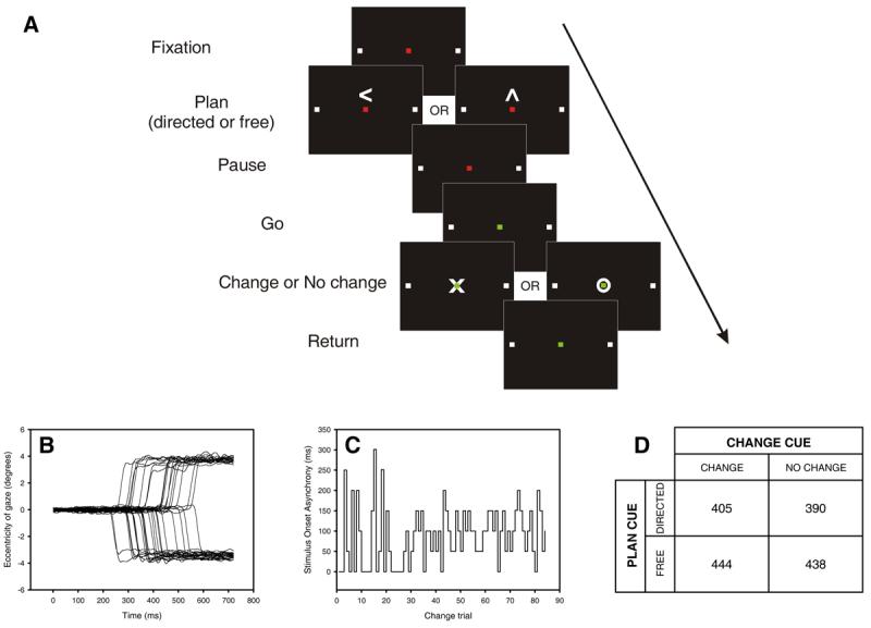
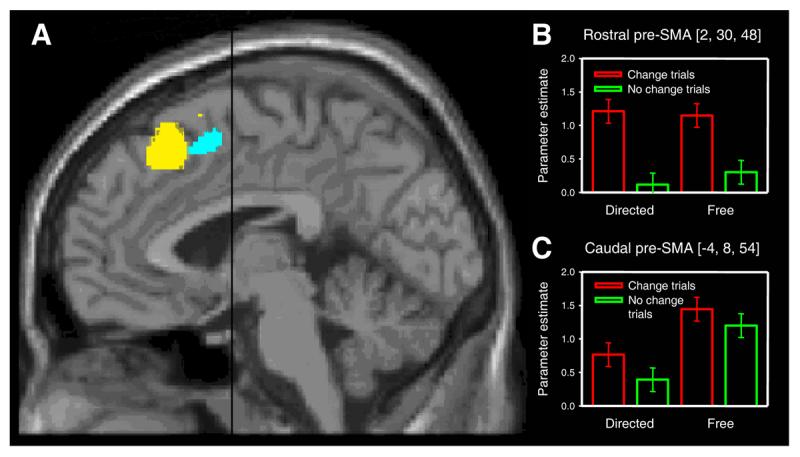
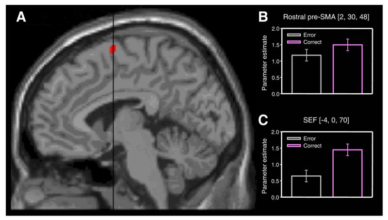
Controversy surrounds the role of human medial frontal cortex in controlling actions. Although damage to this area leads to severe difficulties in spontaneously initiating actions, the precise mechanisms underlying such "volitional" deficits remain to be established. Previous studies have implicated the medial frontal cortex in conflict monitoring and the control of voluntary action, suggesting that these key processes are functionally related or share neural substrates. Here, we combine a novel behavioral paradigm with functional imaging of the oculomotor system to reveal, for the first time, a functional subdivision of the pre-supplementary motor area (pre-SMA) into anatomically distinct areas that respond exclusively to either volition or conflict. We also demonstrate that activity in the supplementary eye field (SEF) distinguishes between success and failure in changing voluntary action plans during conflict, suggesting a role for the SEF in implementing the resolution of conflicting actions. We propose a functional architecture of human medial frontal cortex that incorporates the generation of action plans and the resolution of conflict.


Figures

Figure 1
Figure 1
A. Temporal sequence of visual cues during each trial (not to scale). While fixating a red square, subjects planned a speeded saccade to one of two targets (white squares) that were either freely chosen (free plan) or specifically indicated (directed plan; here a left plan is illustrated). A change in the fixation cue from red to green (‘go’ cue) signalled the execution of the saccade. A variable interval (SOA) after the ‘go’ cue, and before the saccade was executed, a ‘change’ or ‘no change’ cue instructed subjects either to continue with their plan or to execute a saccade to the opposite target instead. The SOA was modulated on-line to target a 50% success rate in directed change trials. B. Raw saccadic traces from one subject performing the task in the scanner (negative eccentricity indicates leftward displacement). Data from left directed change trials is shown. C. Plot of stimulus onset asynchrony (SOA) for directed change trials performed by one subject. After each direct change trial the SOA was automatically increased or decreased by 50 ms depending on whether the subject succeeded or failed in changing plan[40]. The algorithm sampled randomly from two independent threads starting at 0 and 300 ms. D. Group mean of individual subject median saccadic latencies (ms) for each trial type collected during scanning. At the group level, there was no significant main effect of choice (p = 0.09) or conflict (p = 0.57) on Friedman's test.
Figure 2
Figure 2
Pre-SMA activation associated with changing volitional plan and with free choice. A. Statistical parametric maps showing group main effects of changing plan in rostral pre-SMA (yellow, coordinates 2, 30, 48) and free choice in caudal pre-SMA (cyan, coordinates −4, 8, 54) at a threshold of p < 0.001 uncorrected, superimposed on a MNI standard single subject T1-weighted MRI scan. Black line indicates position of anterior commissure (VCA line). B. Signal change in rostral pre-SMA cluster indexed by the parameter estimates for each of the four main conditions. Note significant conflict-related activity on both free and directed trials. Error bars correspond to 90% confidence intervals. C. Corresponding plot for the caudal pre-SMA cluster showing a main effect of free choice and absence of significant conflict-related activity. Neither rostral nor caudal pre-SMA showed a significant interaction (at a threshold of p < 0.001 uncorrected) between the effects of the two factors (choice and conflict), suggesting that volition does not modulate the activation of the conflict-related area, and conflict does not modulate the activation of the volition-related area.
Figure 3
Figure 3
SEF activation associated with successfully changing plans. A. Comparison of successfully changed versus unsuccessfully changed directed trials reveals activity in the SEF. The statistical parametric map has been thresholded at p < 0.001 uncorrected, and superimposed on a MNI standard single subject T1-weighted MRI scan. Black line indicates position of the anterior commissure (VCA line). B. Signal change in the conflict-responsive rostral pre-SMA cluster (see Fig. 2) indexed by the parameter estimates for unsuccessfully (yellow) and successfully (blue) changed directed trials. Note absence of a significant difference (at p < 0.001 uncorrected). Error bars correspond to 90% confidence intervals. C. Corresponding plot for the SEF cluster showing that activity in this region discriminates between success and failure in changing oculomotor plan.

References

    1. Picard N, Strick PL. Imaging the premotor areas. Curr Opin Neurobiol. 2001;11:663–672. - PubMed
    1. Paus T. Primate anterior cingulate cortex: where motor control, drive and cognition interface. Nat Rev Neurosci. 2001;2:417–424. - PubMed
    1. Botvinick MM, Braver TS, Barch DM, Carter CS, Cohen JD. Conflict monitoring and cognitive control. Psychol Rev. 2001;108:624–652. - PubMed
    1. Rushworth MF, Walton ME, Kennerley SW, Bannerman DM. Action sets and decisions in the medial frontal cortex. Trends Cogn Sci. 2004;8:410–417. - PubMed
    1. Gehring WJ, Fencsik DE. Functions of the medial frontal cortex in the processing of conflict and errors. J Neurosci. 2001;21:9430–9437. - PMC - PubMed

Publication types

LinkOut - more resources