. 2005 Jan 31;168(3):477-88.
doi: 10.1083/jcb.200407113. Epub 2005 Jan 24.

Clathrin- and caveolin-1-independent endocytosis: entry of simian virus 40 into cells devoid of caveolae

Affiliations

Clathrin- and caveolin-1-independent endocytosis: entry of simian virus 40 into cells devoid of caveolae

Eva-Maria Damm et al. J Cell Biol. .

Abstract

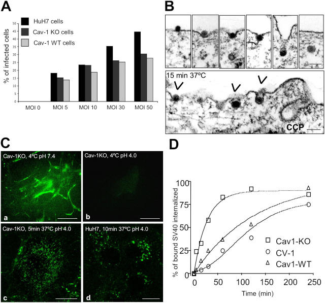
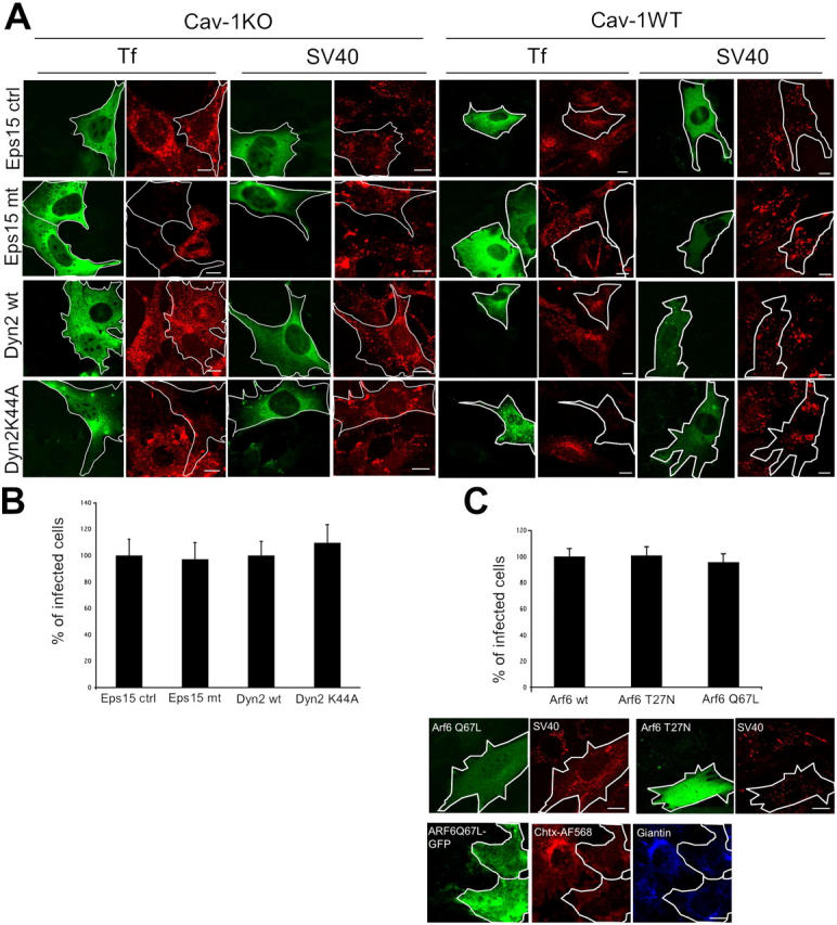
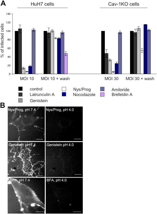
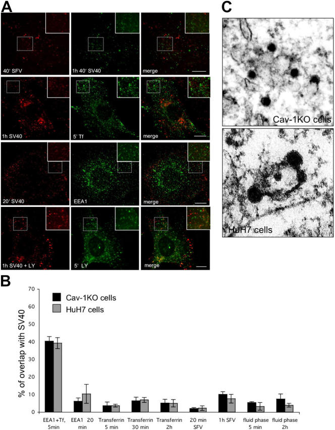
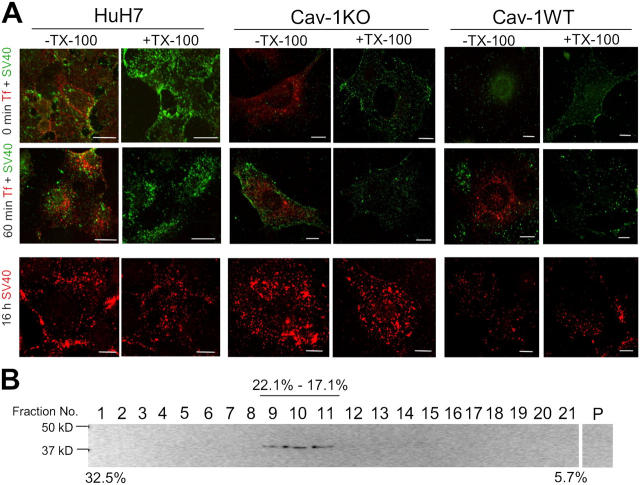
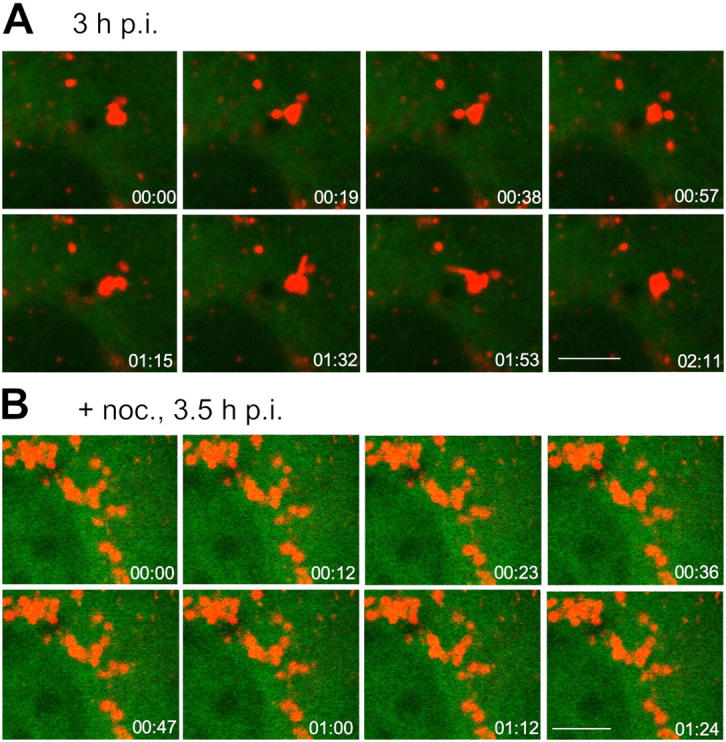
Simian Virus 40 (SV40) has been shown to enter host cells by caveolar endocytosis followed by transport via caveosomes to the endoplasmic reticulum (ER). Using a caveolin-1 (cav-1)-deficient cell line (human hepatoma 7) and embryonic fibroblasts from a cav-1 knockout mouse, we found that in the absence of caveolae, but also in wild-type embryonic fibroblasts, the virus exploits an alternative, cav-1-independent pathway. Internalization was rapid (t1/2 = 20 min) and cholesterol and tyrosine kinase dependent but independent of clathrin, dynamin II, and ARF6. The viruses were internalized in small, tight-fitting vesicles and transported to membrane-bounded, pH-neutral organelles similar to caveosomes but devoid of cav-1 and -2. The viruses were next transferred by microtubule-dependent vesicular transport to the ER, a step that was required for infectivity. Our results revealed the existence of a virus-activated endocytic pathway from the plasma membrane to the ER that involves neither clathrin nor caveolae and that can be activated also in the presence of cav-1.


Figures

Figure 1.
Figure 1.
Cav-1KO and HuH7 cells internalize SV40 and are infected. (A) All cell lines were incubated with different MOIs and analyzed for T-antigen expression after 20 h. (B) Electron micrographs of cav-1KO cells after incubation for 15 min with SV40 at 37°C. Arrowheads point to internalizing virus particles. CCP, clathrin-coated pit. Bars, 100 nm. (C) FITC-labeled SV40 was bound to cav-1KO and HuH7 cells at 4°C and allowed to internalize after shifting to 37°C. Lowering the extracellular pH to 4.0 allowed visualization of only those virus particles that had been internalized. Bars, 10 μm. (D) Internalization of 125I-labeled biotin-SS-SV40 in cav-1WT, cav-1KO, and CV-1 cells. Cells were incubated at 4°C for 2 h and shifted to 37°C in the continuous presence of the virus. At the indicated time points, cells were analyzed for the amount of internalized virus (Pelkmans et al., 2002).
Figure 2.
Figure 2.
Internalization and infection of SV40 occurs independently of Eps15, Dynamin II, and ARF6. Cav-1KO and cav-1WT cells were transfected with Eps15IIIΔ2 (Eps15 ctrl), Eps15 EΔ95/295 (Eps15 mt), Dyn2wt, or Dyn2K44A, all tagged with GFP. (A) After transfection, cells were incubated with AF594-SV40 for 2 h, fixed, and examined in confocal microscopy. AF594-Tf served as a positive control. Representative images are shown. Bars, 10 μm. (B) After transfection, cav-1KO cells were infected with SV40 for 20 h, fixed, and analyzed for infection. Infection in cells expressing the control constructs was set at 100%. Values are given as the mean ± SD. (C) Cav-1KO cells were cotransfected with ARF6 wild type, ARF6 T27N, ARF6 Q67L, and GFP. After transfection, cells were infected with SV40 for 20 h, fixed, and analyzed for infection. Infection in cells expressing the wild-type construct was set at 100%. Values are given as the mean ± SD. Alternatively, cells were incubated with AF594-SV40 for 1.5 h and imaged live. As a positive control, ARF6 Q67L-GFP–transfected cells were incubated with cholera toxin-AF568, fixed, and immunostained with an anti-giantin antibody (blue). Bars, 10 μm.
Figure 3.
Figure 3.
Caveolae- and clathrin-independent SV40 infection requires cholesterol, tyrosine kinases, and a functional microtubule cytoskeleton. (A) HuH7 and cav-1KO cells were either left untreated (control) or pretreated with the indicated drugs and incubated with SV40 for 2 h in the presence of drugs. Virus was removed and cells were further incubated either in the presence of drugs or as a control after drug washout (MOI 10 + wash in HuH7 and MOI 30 + wash in cav-1KO cells). 20 h after infection, cells were analyzed for expression of T-antigen. Values are given as the mean ± SD. (B) Cav-1KO cells were pretreated with nystatin/progesterone, genistein, or BFA and incubated with FITC-SV40 for 1 h. Fluorescence of not internalized viruses was quenched by lowering the extracellular pH to 4. Bars, 10 μm.
Figure 4.
Figure 4.
SV40-containing organelles are distinct from organelles of the classical endosomal pathway. (A) Cav-1KO cells were incubated with AF488-SV40 or AF594-SV40 and the endosomal markers AF594-SFV (first row) or AF488-Tf (second row) for the indicated times, fixed, and analyzed by confocal microscopy. SV40-carrying vesicles did not contain markers specific for clathrin-mediated endocytosis. The same was shown by immunofluorescence of fixed cav-1KO cells (third row) incubated with AF594-SV40 and stained with the early endosomal marker EEA1 (green). In cav-1KO cells incubated with AF594-SV40 and the fluid phase marker Lucifer yellow (LY), only a minor portion of SV40-carrying vesicles contained LY (fourth row). Bars, 10 μm. (B) Quantification of colocalization with endosomal and fluid phase markers in cav-1KO and HuH7 cells after various times. As a control, the first two bars show overlap between Tf and EEA1. Values are given as the mean ± SD. (C) Thin section electron micrographs of cav-1KO and HuH7 cells showing virus particles in intermediate organelles that resemble caveosomes.
Figure 5.
Figure 5.
Association of SV40 with detergent-insoluble membranes. (A) Internalized SV40 but not Tf is resistant to Triton X-100 extraction at 4°C. AF488-SV40 and AF568-Tf were bound to HuH7 (left), cav-1KO (middle), and cav-1WT cells (right) on ice and allowed to internalize for the indicated times. The cells were washed, extracted for 5 min on ice with 1% Triton X-100, and fixed. Bars, 10 μm. (B) SV40 associates and internalizes with DRMs in cav-1–deficient cells. Cav-1KO cells were incubated with SV40 for 30 min at 37°C before cell lysis and extraction with 1% Triton X-100 at 4°C. Samples were floated in a linear sucrose gradient, and fractions were collected and analyzed by SDS-PAGE and immunoblotted against SV40.
Figure 6.
Figure 6.
Endocytosis of SV40 into cav-1–deficient cells results in the accumulation of virus in cytosolic organelles. Cav-1KO cells were transfected with YFP-α-tubulin and incubated with AF594-SV40 for the indicated times. Before virus addition, cells had either been treated with nocodazole (B) or left untreated (A). Movies were recorded by confocal live microscopy of which a series of frames is shown. In nocodazole-treated cells, virus accumulated in caveosome-like organelles, which remained stationary. In A, a series of frames of untreated cells shows the formation of virus-containing transport vesicles from a larger cytosolic organelle. Bars, 5 μm.
Figure 7.
Figure 7.
18 h after internalization, SV40 accumulates in the smooth ER. (A) Cav-1KO cells were incubated with AF594-SV40, fixed after 18 h, stained in indirect immunofluorescence with a Golgi marker (ManII, mannosidase II), an ER marker (CNX, Calnexin), and a marker for the smooth ER (Syn17, syntaxin 17), and analyzed by confocal microscopy. Histograms show colocalization of SV40 with the respective markers over time. Values are given as the mean ± SD. Bars, 10 μm. (B) Thin section electron micrographs of cav-1KO cells infected with SV40 for 18 h showing virus accumulations in tubulo-reticular out-growths of the smooth ER.
Figure 8.
Figure 8.
Cav-1 retransfection shifts the endocytic process back to caveosomes. (A) Confocal fluorescence microscopy of live cav-1KO and cav-1WT cells retransfected with cav-1-mRFP and infected with AF488-SV40. 10 min after warming (a and d), only a portion of AF488-SV40 colocalized with cav-1-mRFP (arrowheads) as quantified in panels c and f. In both cell types, viruses merged with caveosomes (b and e) at later time points. Values are given as the mean ± SD. Bars, 10 μm. (B) Series of frames taken from Video 6. Arrowheads point toward viruses that are subsequently internalized as shown by the dotted circle in the last frame. (arrow) Note that this particle colocalizes with cav-1 and was there in the previous frames. Bars, 5 μm.

References

    1. Anderson, H.A., Y. Chen, and L.C. Norkin. 1996. Bound simian virus 40 translocates to caveolin-enriched membrane domains, and its entry is inhibited by drugs that selectively disrupt caveolae. Mol. Biol. Cell. 7:1825–1834. - PMC - PubMed
    1. Anderson, R.G. 1998. The caveolae membrane system. Annu. Rev. Biochem. 67:199–225. - PubMed
    1. Arnaoutova, I., C.L. Jackson, O.S. Al-Awar, J.G. Donaldson, and Y.P. Loh. 2003. Recycling of Raft-associated prohormone sorting receptor carboxypeptidase E requires interaction with ARF6. Mol. Biol. Cell. 14:4448–4457. - PMC - PubMed
    1. Benlimame, N., P.U. Le, and I.R. Nabi. 1998. Localization of autocrine motility factor receptor to caveolae and clathrin-independent internalization of its ligand to smooth endoplasmic reticulum. Mol. Biol. Cell. 9:1773–1786. - PMC - PubMed
    1. Benmerah, A., M. Bayrou, N. Cerf-Bensussan, and A. Dautry-Varsat. 1999. Inhibition of clathrin-coated pit assembly by an Eps15 mutant. J. Cell Sci. 112:1303–1311. - PubMed

Publication types

MeSH terms

Substances