Comparative Study
. 2005 Feb 1;102(5):1773-8.
doi: 10.1073/pnas.0409227102. Epub 2005 Jan 24.

Structural and biochemical studies identify tobacco SABP2 as a methyl salicylate esterase and implicate it in plant innate immunity

Affiliations
Comparative Study

Structural and biochemical studies identify tobacco SABP2 as a methyl salicylate esterase and implicate it in plant innate immunity

Farhad Forouhar et al. Proc Natl Acad Sci U S A. .

Abstract

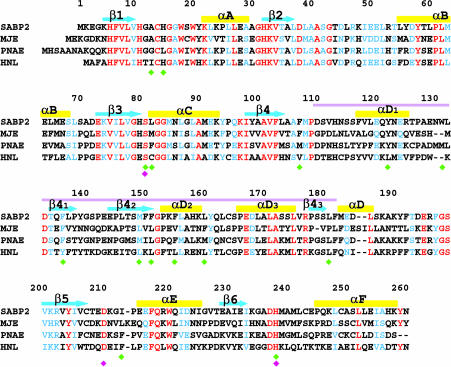
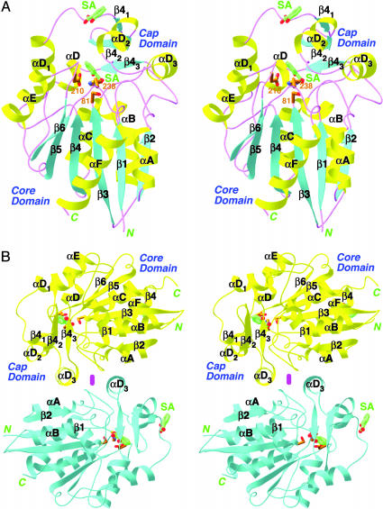
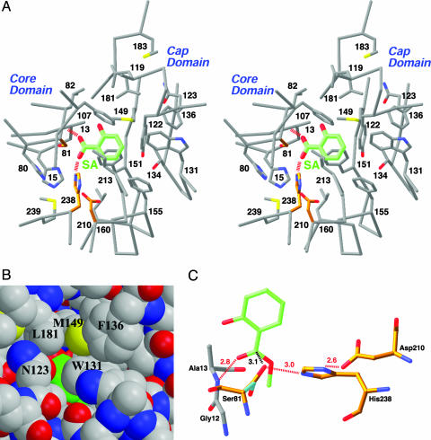
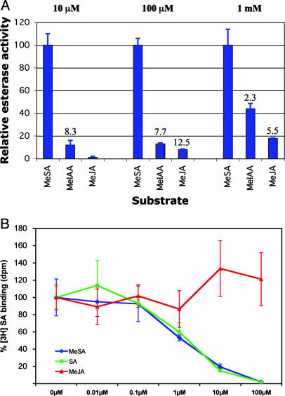
Salicylic acid (SA) is a critical signal for the activation of plant defense responses against pathogen infections. We recently identified SA-binding protein 2 (SABP2) from tobacco as a protein that displays high affinity for SA and plays a crucial role in the activation of systemic acquired resistance to plant pathogens. Here we report the crystal structures of SABP2, alone and in complex with SA at up to 2.1-A resolution. The structures confirm that SABP2 is a member of the alpha/beta hydrolase superfamily of enzymes, with Ser-81, His-238, and Asp-210 as the catalytic triad. SA is bound in the active site and is completely shielded from the solvent, consistent with the high affinity of this compound for SABP2. Our biochemical studies reveal that SABP2 has strong esterase activity with methyl salicylate as the substrate, and that SA is a potent product inhibitor of this catalysis. Modeling of SABP2 with MeSA in the active site is consistent with all these biochemical observations. Our results suggest that SABP2 may be required to convert MeSA to SA as part of the signal transduction pathways that activate systemic acquired resistance and perhaps local defense responses as well.


Figures

Fig. 1.
Fig. 1.
Sequence comparison of tobacco SABP2 with the most similar proteins of known function. These include tomato MeJA esterase (MJE), Rauvolfia serpentina polyneuridine aldehyde esterase (PNAE), and Brazil nut HNL. Identical residues are shown in red and similar residues in blue. The cyan arrows and yellow bars identify the secondary structure elements. The purple line marks the cap domain. The catalytic triad residues are indicated with the magenta diamond, and residues that contact SA are indicated by green diamonds. See Fig. 5 for the GenBank accession nos. of these sequences and for an alignment with additional sequences.
Fig. 2.
Fig. 2.
Structure of SABP2 in complex with SA. (A) Stereoview of the SABP2 monomer in complex with SA. The core and cap domains are labeled. The secondary structure elements, α helices, β strands, and loops are colored in yellow, cyan, and magenta, respectively. SA (in green for carbon atoms) is located in the active site as well as another site on the surface of the enzyme. This second surface-binding site may not be physiologically relevant. (B) Dimer of SABP2. The two monomers are colored in yellow and cyan, respectively. The 2-fold axis of the dimer is indicated with the magenta oval [produced with ribbons (39)].
Fig. 3.
Fig. 3.
The active site of SABP2 and the binding mode of SA. (A) Stereoview of the active site of SABP2 in complex with SA. The catalytic triad residues (Ser-81, His-238, and Asp-210) are shown in gold. The hydrogen bonds from the carboxylate group of SA are shown as red dashed lines. (B) Model of SABP2 in complex with SA, showing that the SA molecule (in green for carbon atoms) is shielded from the solvent in the active site. (C) Model of the binding mode of the MeSA substrate (green) to SABP2. The side chain of the catalytic Ser-81 residue assumes a different conformation for catalysis (cyan and gold in complex with SA and MeSA, respectively). The hydrogen bonds are indicated in dashed lines in red, and the distance between Ser-81, and the MeSA carboxylate carbon is indicated in black.
Fig. 4.
Fig. 4.
Comparisons of methyl esterase activities and binding affinities of SABP2. (A) Relative methyl esterase activity of SABP2 with MeSA, MeIAA, and MeJA substrates at three different concentrations (10 μM, 100 μM, and 1 mM). The activity with MeSA at each of the substrate concentrations was set at 100%. The activity ratios between MeSA and the other substrates are indicated. All results are the average of three independent experiments. (B) SABP2 binds MeSA but not MeJA. Binding of [3H]SA by SABP2 in the absence of any competitor (SA, MeSA, or MeJA) was set to 100%. MeSA (blue) competes with [3H]SA for binding to SABP2 with the same potency as SA (green), whereas MeJA (red) does not compete for binding. Results (±SD) from competition-binding assays are presented as an average of three replicate binding reactions. The binding experiments were repeated three times with similar results.

References

    1. Dangl, J. L. & Jones, J. D. G. (2001) Nature 411, 826–833. - PubMed
    1. Holt, B. F., III, Hubert, D. A. & Dangl, J. L. (2003) Curr. Opin. Immunol. 15, 20–25. - PubMed
    1. Cohn, J., Sessa, G. & Martin, G. B. (2001) Curr. Opin. Immunol. 13, 55–62. - PubMed
    1. Hammond-Kosack, K. E. & Jones, J. D. G. (1996) Plant Cell 8, 1773–1791. - PMC - PubMed
    1. Dempsey, D., Shah, J. & Klessig, D. F. (1999) Crit. Rev. Plant Sci. 18, 547–575.

Publication types