. 2005 Feb 1;102(5):1584-9.
doi: 10.1073/pnas.0409191102. Epub 2005 Jan 24.

Nibrin functions in Ig class-switch recombination

Affiliations

Nibrin functions in Ig class-switch recombination

Sven Kracker et al. Proc Natl Acad Sci U S A. .

Abstract

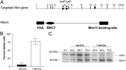
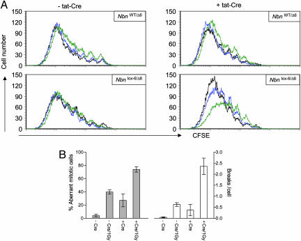
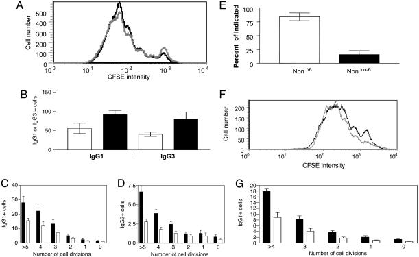
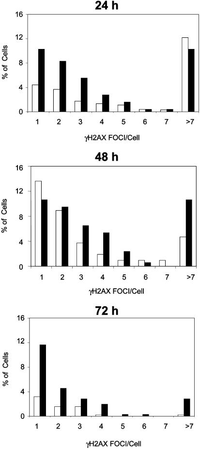
Nijmegen breakage syndrome (NBS) is a rare autosomal recessive disorder characterized by predisposition to hematopoietic malignancy, cell-cycle checkpoint defects, and ionizing radiation sensitivity. NBS is caused by a hypomorphic mutation of the NBS1 gene, encoding nibrin, which forms a protein complex with Mre11 and Rad50, both involved in DNA repair. Nibrin localizes to chromosomal sites of class switching, and B cells from NBS patients show an enhanced presence of microhomologies at the sites of switch recombination. Because nibrin is crucial for embryonic survival, direct demonstration by targeted deletion that nibrin functions in class switch recombination has been lacking. Here, we show by cell-type-specific conditional inactivation of Nbn, the murine homologue of NBS1, that nibrin plays a role in the repair of gamma-irradiation damage, maintenance of chromosomal stability, and the recombination of Ig constant region genes in B lymphocytes.


Figures

Fig. 1.
Fig. 1.
Generation of nibrin-deficient B lymphocytes. (A) Schematic representation of the targeted Nbn gene locus and the protein structure of nibrin. (B) Efficiency of tat-Cre-mediated Nbn exon 6 deletion. Nested single-cell PCR detects the deleted allele and the floxed exon 6 Nbn allele. Data represent the mean and SD of five individual cultures. (C) Expression of nibrin and Mre11 proteins in populations of LPS/IL4-activated Nbnlox-6/Δ6 B lymphocytes, not treated or treated with tat-Cre in vitro, at the indicated time points after activation. Cell extracts of 5 × 10⁵ cells each were submitted to Western blot analysis, as described in Materials and Methods.
Fig. 2.
Fig. 2.
Radiation sensitivity and chromosomal instability of nibrin-deficient B lymphocytes. (A) Radiation sensitivity of Nbnlox-66 and NbnWT6 B lymphocytes after deletion of exon 6 of Nbn by tat-Cre in vitro. Cells were labeled with CFSE and activated with LPS/IL4 for 3 days. Cells were then not irradiated intentionally (black line), or γ-irradiated with 1.5 Gy (blue) or 3.6 Gy (green line). Two days later, proliferation and survival of the cells was analyzed cytometrically. Proliferative history of surviving cells was determined according to loss of CFSE label and numbers of surviving cells relative to reference beads (TrueCount, Becton Dickinson), standardizing the numbers of recorded viable cells not stained for propidium iodide. (B) Chromosomal instability of LPS/IL4-activated Nbnlox-6/Δ6 B lymphocytes after treatment with tat-Cre in vitro. On day 3 of culture, metaphase spreads were prepared from cells 3 h after γ-irradiation (1 Gy). Metaphases were Giemsa-stained and scored for aberrant chromosome numbers and apparent chromosomal breaks (-Cre/-Gy, 102 metaphases analyzed; -Cre/+Gy, 59 metaphases; +Cre/-Gy, 236 metaphases; +Cre/+Gy, 138 metaphases).
Fig. 3.
Fig. 3.
Antibody class-switch recombination in B lymphocytes conditionally inactivated for Nbn in vitro or in vivo. (A) Proliferation according to loss of CFSE label of Nbnlox-66 B lymphocytes analyzed on day 3 of LPS/IL-4 activation, and treated with tat-Cre (solid line) or not treated (shaded line) in vitro, before the onset of activation. Data shown are from a representative experiment. (B) Comparison of Nbnlox-66 (white bars) and NbnWT/lox-6 (black bars) B cells switched to IgG1 upon activation by LPS/IL-4, or switched to IgG3 upon activation by LPS, in tat-Cre treated relative to cultures not treated with tat-Cre. Data represent the mean and SD of individual LPS (n = 6 for Nbnlox-6/Δ6 and n = 4 NbnWT/lox-6)orLPS/IL4 cultures (n = 7 for Nbnlox-6/Δ6 and n = 5 NbnWT/lox-6; P < 0.01, Mann–Whitney U test). Flow cytometric analysis of surface IgG1 (C) and surface IgG3 expression by CFSE-labeled Nbnlox-6/Δ6 B lymphocytes (D) treated with tat-Cre (white bars) or not treated (black bars), after 3 days of LPS/IL-4 and LPS stimulation, respectively. The frequencies of IgG1- or IgG3-expressing cells are shown separately for each population that has undergone the indicated number of cell divisions since the onset of activation, according to CFSE staining. Data represent the mean and SE of four individual LPS/IL4 or LPS cultures. (E) Efficiency of depletion of exon 6 of Nbn in vivo, in CD19-Cre Nbnlox-6/lox-6 B lymphocytes, detected by quantitative PCR specific for the deleted and nondeleted floxed Nbn alleles. (Data from three mice are represented.) (F) Proliferation of CD19-Cre Nbnlox-6/lox-6 (solid line) and CD19-Cre Nbnlox-6/WT B lymphocytes (shaded line) after activation with LPS for 3 days, according to loss of CFSE label. (G) Flow cytometric analysis of surface IgG1 expression on CFSE-labeled CD19-Cre Nbnlox-6/lox-6 (white bars) and CD19-Cre Nbnlox-6/WT B lymphocytes (black bars) stimulated with LPS/IL-4 for 3 days. The frequency of IgG1+ cells is shown for each population that had undergone the indicated number of cell divisions since the onset of activation, according to loss of CFSE label. Data represent the mean and SE of four individual LPS/IL4 cultures.
Fig. 4.
Fig. 4.
Generation and maintenance of γ-H2AX foci in conditionally Nbn-inactivated, activated B lymphocytes. γ-H2AX foci were stained with a specific antibody in CD19-Cre Nbnlox6/lox6 (black bars) and CD19-Cre Nbnlox6/WT B lymphocytes (white bars) after 24, 48, and 72 h of LPS/IL-4 activation. The samples were double-blinded and foci were scored by fluorescence microscopy. (WT/24 h, 976 cells; WT/48 h, 213 cells; WT/72 h, 379 cells; lox6/24 h, 253 cells; lox6/48 h, 169 cells; lox6/72 h, 352 cells.)

References

    1. Varon, R., Vissinga, C., Platzer, M., Cerosaletti, K. M., Chrzanowska, K. H., Saar, K., Beckmann, G., Seemanova, E., Cooper, P. R., Nowak, N. J., et al. (1998) Cell 93, 467-476. - PubMed
    1. Tauchi, H., Kobayashi, J., Morishima, K., van Gent, D. C., Shiraishi, T., Verkaik, N. S., vanHeems, D., Ito, E., Nakamura, A., Sonoda, E., et al. (2002) Nature 420, 93-98. - PubMed
    1. Zhu, J., Petersen, S., Tessarollo, L. & Nussenzweig, A. (2001) Curr. Biol. 11, 105-109. - PubMed
    1. Xiao, Y. & Weaver, D. T. (1997) Nucleic Acids Res. 25, 2985-2991. - PMC - PubMed
    1. Luo, G., Yao, M. S., Bender, C. F., Mills, M., Bladl, A. R., Bradley, A. & Petrini, J. H. (1999) Proc. Natl. Acad. Sci. USA 96, 7376-7381. - PMC - PubMed

Publication types