Reduced medial temporal lobe N-acetylaspartate in cognitively impaired but nondemented patients
- PMID: 15668426
- PMCID: PMC1851679
- DOI: 10.1212/01.WNL.0000149638.45635.FF
Reduced medial temporal lobe N-acetylaspartate in cognitively impaired but nondemented patients
Abstract
Background: N-acetylaspartate (NAA) in the medial temporal lobe (MTL) and parietal lobe gray matter (GM) is diminished in Alzheimer disease (AD). Because NAA is considered a marker of neuronal integrity, reduced medial temporal and parietal lobe NAA could be an early indication of dementia-related pathology in elderly individuals.
Objectives: 1) To determine whether cognitively impaired but nondemented (CIND) elderly individuals exhibit a similar pattern of reduced medial temporal and parietal lobe NAA as AD patients. 2) To compare regional NAA patterns, hippocampal and neocortical gray matter (GM) volumes in CIND patients who remained cognitively stable and those who became demented over 3.6 years of follow-up. 3) To examine the relationship between memory performance, medial temporal lobe NAA, and hippocampal volume.
Methods: Seventeen CIND, 24 AD, and 24 cognitively normal subjects were studied using MRSI and MRI.
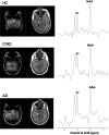
Results: Relative to controls, CIND patients had reduced MTL NAA (19 to 21%, p = 0.005), hippocampal (11 to 14%, p < or = 0.04), and neocortical GM (5%, p = 0.05) volumes. CIND patients who later became demented had less MTL NAA (26%, p = 0.01), hippocampal (17 to 23%, p < or = 0.05), and neocortical GM (13%, p = 0.02) volumes than controls, but there were no significant differences between stable CIND patients and controls. MTL NAA in combination with hippocampal volume improved discrimination of CIND and controls over hippocampal volume alone. In AD and CIND patients, decreased MTL NAA correlated significantly with impaired memory performance.
Conclusion: Reduced medial temporal lobe N-acetylaspartate, together with reduced hippocampal and neocortical gray matter volumes, may be early indications of dementia-related pathology in subjects at high risk for developing dementia.
Figures

References
-
- Laukka EJ, Jones S, Small BJ, Fratiglioni L, Backman L. Similar patterns of cognitive deficits in the preclinical phases of vascular dementia and Alzheimer's disease. J Int Neuropsychol Soc. 2004;10:382–391. - PubMed
-
- Valenzuela MJ, Sachdev P. Magnetic resonance spectroscopy in AD. Neurology. 2001;56:592–598. - PubMed
-
- Crook TH, Bartus RT, Ferris SH, Whitehouse P, Cohen GD, Gershon S. Age-associated memory impairment: proposed diagnostic criteria and measures of clinical change: report of a National Institute of Mental Health Work Group. Dev Neuropsychol. 1986;2:261–276.
-
- Petersen RC, Smith GE, Waring SC, Ivnik RJ, Tangalos EG, Kokmen E. Mild cognitive impairment: clinical characterization and outcome. Arch Neurol. 1999;56:303–308. - PubMed
-
- Parnetti L, Lowenthal DT, Presciutti O, et al. 1H-MRS, MRI-based hippocampal volumetry, and 99mTc-HMPAO-SPECT in normal aging, age-associated memory impairment, and probable Alzheimer's disease. J Am Geriatr Soc. 1996;44:133–138. - PubMed
Publication types
MeSH terms
Substances
Grants and funding
LinkOut - more resources
Full Text Sources
