. 2005 Feb;115(2):339-47.
doi: 10.1172/JCI23183.

Transcriptional activation of integrin beta6 during the epithelial-mesenchymal transition defines a novel prognostic indicator of aggressive colon carcinoma

Affiliations

Transcriptional activation of integrin beta6 during the epithelial-mesenchymal transition defines a novel prognostic indicator of aggressive colon carcinoma

Richard C Bates et al. J Clin Invest. 2005 Feb.

Abstract

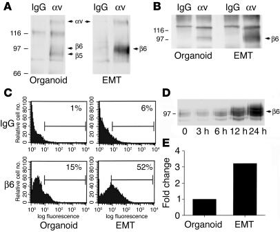
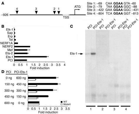
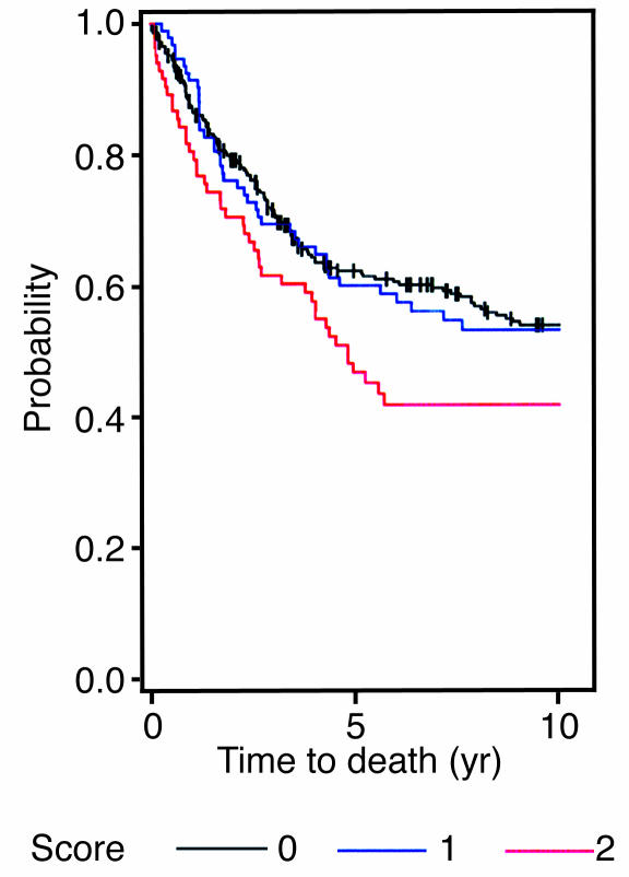
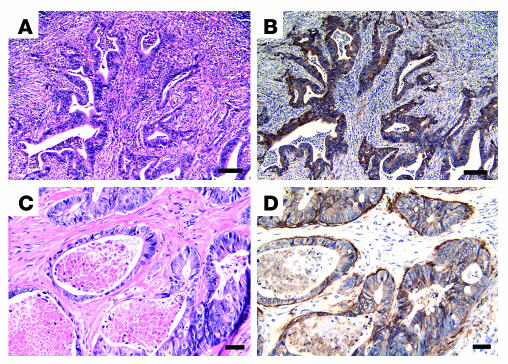
We used a spheroid model of colon carcinoma to analyze integrin dynamics as a function of the epithelial-mesenchymal transition (EMT), a process that provides a paradigm for understanding how carcinoma cells acquire a more aggressive phenotype. This EMT involves transcriptional activation of the beta6 integrin subunit and a consequent induction of alphavbeta6 expression. This integrin enhances the tumorigenic properties of colon carcinoma, including activation of autocrine TGF-beta and migration on interstitial fibronectin. Importantly, this study validates the clinical relevance of the EMT. Kaplan-Meier analysis of beta6 expression in 488 colorectal carcinomas revealed a striking reduction in median survival time of patients with high beta6 expression. Elevated receptor expression did not simply reflect increasing tumor stage, since log-rank analysis showed a more significant impact on the survival of patients with early-stage, as opposed to late-stage, disease. Cox regression analysis confirmed that this integrin is an independent variable for these tumors. These findings define the alphavbeta6 integrin as an important risk factor for early-stage disease and a novel therapeutic candidate for colorectal cancer.


Figures

Figure 1
Figure 1
Integrin αvβ6 expression increases following EMT. (A) LIM 1863 organoids or cells harvested 24 hours after induction of EMT were surface biotinylated, and the extracts were immunoprecipitated with an mAb directed against integrin αv or control IgG. Relative molecular masses are shown to the left in kDa. Arrows indicate the positions of αv, and its associated chains β5 and β6. (B) Cell extracts were prepared from organoids or from cells 24 hours after EMT, immunoprecipitated as in A, and then immunoblotted with an mAb against the β6 integrin subunit. Relative molecular masses are shown to the left in kDa. (C) LIM 1863 organoids were either disaggregated into single-cell suspensions (Organoid) or harvested 24 hours after induction of the EMT (EMT), and surface expression of β6 was assessed by flow cytometry. A greater–than–3-fold increase in β6 surface expression occurs after the EMT. (D) Cell extracts were prepared over the time course shown after EMT induction and immunoblotted with an anti-β6 antibody to determine the kinetics of upregulation of the receptor. Relative molecular masses are shown to the left in kDa. (E) Integrin β6 mRNA levels were quantified using real-time quantitative PCR in organoid or EMT cultures of LIM 1863 cells. The fold change between treatments (3.2-fold induction following the EMT) is represented graphically.
Figure 2
Figure 2
Transcriptional regulation of β6 by the Ets-1 transcription factor. (A) Schematic of the human integrin β6 promoter. The transcription start site (TSS) and translation start site (ATG) are indicated. Putative Ets-binding sites are shown (triangles), and the 4 corresponding sequences are listed, including location detail. The consensus DNA-binding sequence (GGAA) is shown in bold. (B) Transactivation of the β6 luciferase reporter construct (–926/+208) by a panel of Ets transcription factors compared with the empty mammalian expression plasmid (PCI) in HEK293 cells. The change in luciferase activity is expressed as fold induction compared with PCI. (C) Gel mobility shift assay for Ets-1 binding to putative Ets sites in the β6 promoter. In vitro–translated Ets-1 protein or control extract was used with end-labeled oligonucleotide probes encoding putative Ets sites 1–4, as indicated. (D) Mutational analysis of the β6 promoter. Transactivation of either the wild-type (black bars) or mutant Ets-1–binding site –66/–63 (white bars) β6 luciferase reporter constructs in response to increasing doses of Ets-1 is shown.
Figure 3
Figure 3
Increased αvβ6 expression promotes migration on fibronectin. (A) Chemotactic migration assay of post-EMT LIM 1863 cells for 3 days on untreated control Transwells (Con), or Transwells coated with laminin (Lm) or fibronectin (Fn). Data are expressed as means and SDs of 8 individual fields randomly selected from each well, with each experiment performed in triplicate. *P < 0.05. (B) LIM 1863 cells were subjected to the chemotaxis assay as described, on untreated (Con) or fibronectin-coated Transwells as indicated. Chemotaxis on fibronectin was performed in the absence (–) or presence of function-blocking anti-β6 monoclonal (10D5) or isotype-matched control (IgG) antibody, at a concentration of 100 μg/ml. The anti-β6 antibody reduced chemotaxis back to the levels of the uncoated migration control for these cells. *P < 0.05.
Figure 4
Figure 4
Autocrine TGF-β production and activation by αvβ6 in EMT cells. (A) ELISA was performed to measure secreted TGF-β levels in serum-free media (Media), media after washing (Wash), or overnight culture media from organoid or EMT cultures of LIM 1863 cells (Org, EMT). (B) Reporter and LIM 1863 cells were cultured 16–20 hours and lysed for measurement of luciferase activity. Results are for organoids (Org) and EMT cells (EMT). Addition of function-blocking anti-β6 monoclonal (3G9, 30 μg/ml) or control antibody (Con) is shown at the bottom. Relative luciferase activity (RLU) is the measured activity divided by the activity of the coculture of organoid (control) cells in the presence of function-blocking antibody. *P < 0.001.
Figure 5
Figure 5
The integrin αvβ6 is a marker of EMT in vivo. Integrin β6 (A) or E-cadherin (B) immunostaining of sequential tumor sections derived from LIM 1863 xenografts. E-cadherin expression is prominent in the tumor tissue (T) and absent from the stroma (S). β6 Immunoreactivity is relatively weak in the tumor masses, but it is strongly upregulated in tumor cells invading the stroma, which are also negative for E-cadherin expression (arrows). Scale bar: 50 μm.
Figure 6
Figure 6
Expression of αvβ6 in malignant human colon carcinoma. (AE) Representative β6 immunostaining of normal human colon (A) or malignant colon carcinoma tissue (BE). A negative (B) and a positive (C) tumor sample are shown. β6 Immunostaining is shown for a separate carcinoma sample in D, with tumor cells infiltrating the stroma showing high expression of the receptor. Heterogeneous receptor expression in another primary tumor, with intense upregulation and preferential localization to tumor islets, is illustrated in E. (F) The corresponding negative control for the tumor shown in E. Scale bars: 100 μm (AC, E, and F) and 50 μm (D).
Figure 7
Figure 7
Kaplan-Meier survival analysis using a log-rank test. The samples were grouped according to β6 expression level (score 0, negative; score 1, low expression; score 2, high expression) and analyzed using the log-rank test for overall survival.
Figure 8
Figure 8
In vivo αvβ6 protein expression in human colorectal metastases. Representative H&E (A and C) or β6 immunohistochemistry (B and D) in lymph node (A and B) or liver tissue (C and D) containing human colorectal metastases. β6 Immunoreactivity in both samples is restricted to the metastasized tumor cells. Scale bars: 100 μm (A and B) and 50 μm (C and D).

Similar articles

Cited by

References

    1. Hay ED. An overview of the epithelio-mesenchymal transformation. Acta Anat. 1995;154:8–20. - PubMed
    1. Savagner P. Leaving the neighborhood: molecular mechanisms involved during epithelial-mesenchymal transition. Bioessays. 2001;23:912–923. - PubMed
    1. Theiry JP. Epithelial-mesenchymal transitions in development and pathologies. Curr. Opin. Cell Biol. 2003;15:740–746. - PubMed
    1. Oft M, et al. TGF-beta1 and Ha-Ras collaborate in modulating the phenotypic plasticity and invasiveness of epithelial tumor cells. Genes Dev. 1996;10:2462–2477. - PubMed
    1. Oft M, Heider KH, Berg H. TGFβ signaling is necessary for carcinoma cell invasiveness and metastasis. Curr. Biol. 1998;8:1243–1252. - PubMed

Publication types

MeSH terms