. 2005 Feb;115(2):407-17.
doi: 10.1172/JCI23025.

Human lupus autoantibody-DNA complexes activate DCs through cooperation of CD32 and TLR9

Affiliations

Human lupus autoantibody-DNA complexes activate DCs through cooperation of CD32 and TLR9

Terry K Means et al. J Clin Invest. 2005 Feb.

Abstract

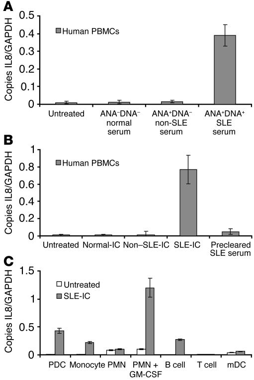
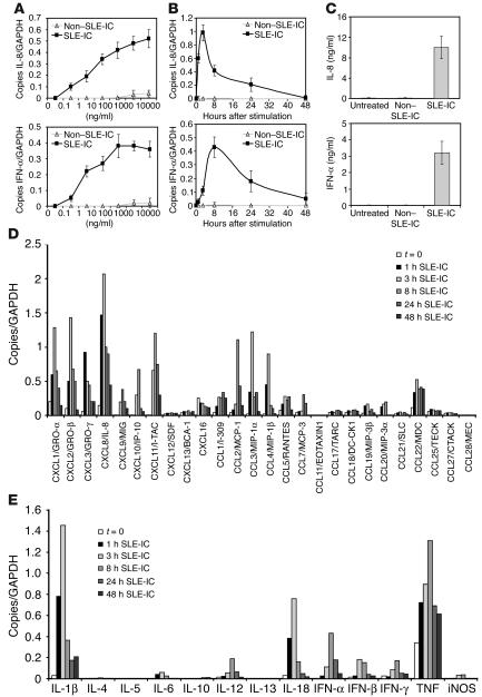
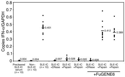
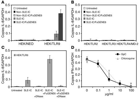
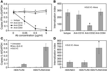
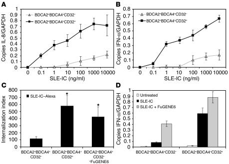
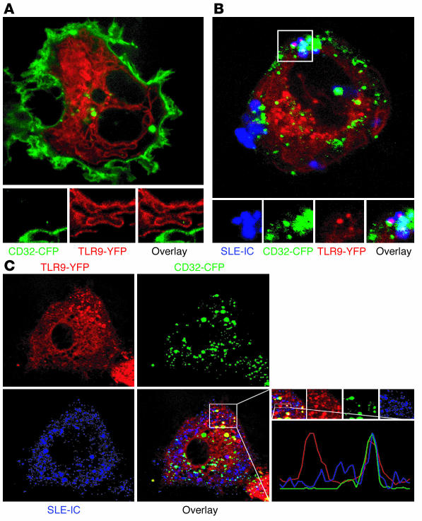
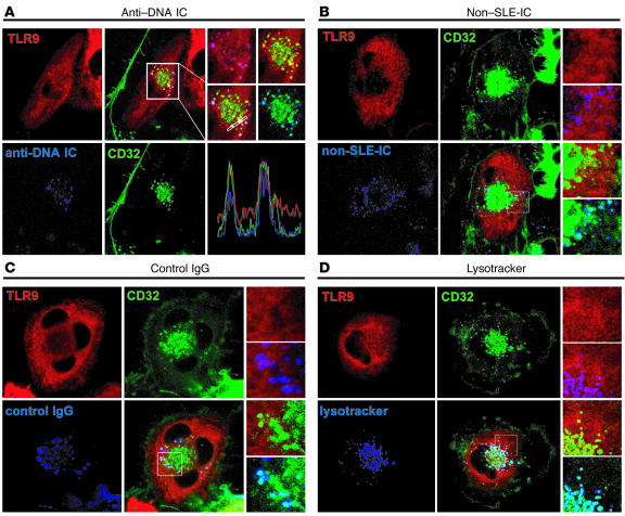
Systemic lupus erythematosus (SLE) is an autoimmune disease characterized by pathogenic autoantibodies against nucleoproteins and DNA. Here we show that DNA-containing immune complexes (ICs) within lupus serum (SLE-ICs), but not protein-containing ICs from other autoimmune rheumatic diseases, stimulates plasmacytoid DCs (PDCs) to produce cytokines and chemokines via a cooperative interaction between Toll-like receptor 9 (TLR9) and FcgammaRIIa (CD32). SLE-ICs transiently colocalized to a subcellular compartment containing CD32 and TLR9, and CD32+, but not CD32-, PDCs internalized and responded to SLE-ICs. Our findings demonstrate a novel functional interaction between Fc receptors and TLRs, defining a pathway in which CD32 delivers SLE-ICs to intracellular lysosomes containing TLR9, inducing a signaling cascade leading to PDC activation. These data demonstrate that endogenous DNA-containing autoantibody complexes found in the serum of patients with SLE activate the innate immune system and suggest a novel mechanism whereby these ICs contribute to the pathogenesis of this autoimmune disease.


Figures

Figure 1
Figure 1
Purified anti-DNA–containing ICs in SLE serum induce cellular activation. (A) Normal human PBMCs were isolated and stimulated with 20% serum isolated from normal patients (ANADNA), non-SLE patients (ANA+DNA, 4 Sjogren syndrome and 4 rheumatoid arthritis patients), or 5 SLE patients (ANA+DNA+). After 8 hours of stimulation, the cells were harvested for RNA. (B) PBMCs were stimulated for 8 hours with 100 ng/ml of ICs purified from the serum samples in A. Some PBMCs were stimulated with a 20% serum equivalent of protein G precleared SLE serum. (C) Purified subsets of white blood cells were stimulated with 100 ng/ml SLE-ICs for 8 hours. Expression of IL-8 was determined by QPCR and depicted as the number of copies of mRNA per copies of the control mRNA GAPDH. Error bars indicate standard deviation of triplicate measurements. Data are representative of 4 similar experiments.
Figure 2
Figure 2
ICs in SLE serum activate cytokine and chemokine production in human PDCs. (A) Normal human PDCs were isolated and stimulated at the indicated doses of purified non–SLE-ICs or SLE-ICs. After 8 hours of stimulation, the cells were harvested for RNA. (B) Normal human PDCs were stimulated with 100 ng/ml of non–SLE-ICs or SLE-ICs. Cells were harvested at the indicated time points for RNA. Expression of IL-8 and IFN-α was determined by QPCR and depicted as the number of copies of mRNA per copies of the control mRNA GAPDH. (C) Supernatants of the stimulated cells were collected after the cells were removed by centrifugation. ELISA was performed to detect human IL-8 and IFN-α at the 24-hour time point. Error bars indicate standard deviation of triplicate measurements. Expression of chemokines (D) and cytokines (E) was quantified by QPCR using total RNA isolated from PDCs (1 × 10⁵ cells) stimulated with 100 ng/ml SLE-ICs for 1, 3, 8, 24, or 48 hours. Data are representative of 4 similar experiments conducted using 4 different donors.
Figure 3
Figure 3
Both the DNA and antibody components of SLE-ICs are necessary for activation of PDCs. Total RNA was isolated from normal human PDCs (1 × 10⁵ cells) stimulated with 100 ng/ml of non–SLE-ICs, SLE-ICs, or SLE-ICs pretreated with immobilized DNase, papain, or pepsin. Some ICs were pretreated with 5 μl of FuGENE6 for 15 minutes prior to stimulation. Expression of IFN-α was determined by QPCR and is depicted as the number of copies of mRNA per copies of the control mRNA GAPDH.
Figure 4
Figure 4
TLR9 confers responsiveness to ICs in SLE serum. (A and B) Total RNA was isolated from HEK cells stably transfected with TLR9, TLR2, TLR3, or TLR4/MD-2 (1 × 10⁶ cells) stimulated with 100 ng/ml non–SLE-ICs or SLE-ICs in the presence or absence of 10 μl of FuGENE6 for 3 hours. NEO, neomycin. (C) Total RNA was isolated from HEK/TLR9 cells stimulated with 100 ng/ml SLE-ICs in the presence or absence of 10 μl FuGENE6 and/or pretreated with DNase. Expression of IL-8 was determined by QPCR and is depicted as the number of copies of mRNA per copies of the control mRNA GAPDH. (D) Human PDCs were stimulated with 100 ng/ml SLE-ICs in the presence or absence of increasing doses of chloroquine or inhibitory synthetic GpC oligonucleotide.
Figure 5
Figure 5
Human CD32 is required for PDC activation and internalization of SLE-ICs. (A) Total RNA was isolated from normal human PDCs stimulated with 100 ng/ml SLE-ICs for 8 hours in the absence or presence of increasing concentrations (0.05, 0.5, 5 μg/ml) of antibodies against CD16, CD32, or CD64. Expression of IFN-α was determined by QPCR and depicted as the number of copies of mRNA per copies of the control mRNA GAPDH. (B and D) Alexa Fluor 633–conjugated SLE-ICs (SLE-IC–Alexa) were incubated with normal human PDCs, HEK/TLR9 cells, or HEK/TLR9/CD32 cells at a ratio of 10:1 for 15 minutes at 37–C; and in the presence or absence of 5 μg/ml of neutralizing antibodies against CD16, CD32, or CD64. Internalization was measured by fluorescent microscopy. Data are presented as the number of internalized complexes per 100 cells × 100. *P < 0.01 versus isotype-treated or control, Student’s t test. Error bars indicate standard deviation of triplicate samples. (C) Total RNA was isolated from HEK/TLR9 or HEK/TLR9/CD32 cells stimulated with 100 ng/ml non–SLE-ICs or SLE-ICs for 3 hours. Expression of IL-8 was determined by QPCR.
Figure 6
Figure 6
CD32+ PDCs internalize and respond to SLE-ICs. (A and B) PDCs from normal donors were isolated on a MoFlo cell sorter using fluorescently labeled antibodies against BDCA2, BDCA4, and CD32. The CD32 and CD32+ PDC subsets were stimulated with the indicated concentrations of SLE-ICs. Cells were harvested at 3 and 8 hours, and expression of IL-8 (A) and IFN-α (B) was determined by QPCR. (C) Alexa Fluor 633–conjugated SLE-ICs in the presence or absence of 10 μl FuGENE6 were incubated with CD32+ and CD32 PDCs at a ratio of 10:1 for 15 minutes at 37–C. Internalization was measured by fluorescent microscopy. Data are presented as the number of internalized complexes per 100 cells × 100. *P < 0.01 versus control, Student’s t test. Error bars indicate standard deviation of triplicate samples. (D) Total RNA was isolated from PDCs stimulated with 100 ng/ml SLE-ICs in the presence or absence of 10 μl of FuGENE6. Expression of IFN-α was determined by QPCR and is depicted as the number of copies of mRNA per copies of the control mRNA GAPDH.
Figure 7
Figure 7
SLE-ICs associate with TLR9 and CD32. U373 cells coexpressing YFP-tagged TLR9 (red) and CFP-tagged CD32 (green) were left untreated (A) or incubated with Alexa Fluor 633–conjugated SLE-ICs (blue) for 5 minutes (B and C), and living cells were imaged by confocal microscopy.
Figure 8
Figure 8
CD32 delivers anti-DNA ICs to TLR9-containing lysosomes. U373 cells coexpressing YFP-tagged TLR9 (red) and CFP-tagged CD32 (green) were incubated with Alexa Fluor 633–conjugated (blue) anti-DNA ICs (A), Alexa Fluor 633–conjugated non–SLE-ICs (B), or Alexa Fluor 633–conjugated IgG (C) for 5 minutes, and living cells were imaged by confocal microscopy. (D) Cells expressing TLR9-CFP (red) and CD32-CFP (green) were incubated with lysotracker (blue).

Similar articles

Cited by

References

    1. Mok CC, Lau CS. Pathogenesis of systemic lupus erythematosus. J. Clin. Pathol. 2003;56:481–490. - PMC - PubMed
    1. Lawrence RC, et al. Estimates of the prevalence of arthritis and selected musculoskeletal disorders in the United States. Arthritis Rheum. 1998;41:778–799. - PubMed
    1. Bootsma H, et al. Anti-double stranded DNA antibodies in systemic lupus erythematosus: detection and clinical relevance of IgM-class antibodies. Scand. J. Rheumatol. 1996;25:352–359. - PubMed
    1. Arbuckle MR, et al. Development of autoantibodies before the clinical onset of systemic lupus erythematosus. N. Engl. J. Med. 2003;349:1526–1533. - PubMed
    1. ter Borg EJ, Horst G, Hummel EJ, Limburg PC, Kallenberg CG. Measurement of increases in anti-double-stranded DNA antibody levels as a predictor of disease exacerbation in systemic lupus erythematosus. A long-term, prospective study. Arthritis Rheum. 1990;33:634–643. - PubMed

Publication types

MeSH terms