

Expression of STAT1 and STAT2 in malignant melanoma does not correlate with response to interferon-alpha adjuvant therapy
- PMID: 15668815
- PMCID: PMC11032810
- DOI: 10.1007/s00262-004-0649-y
Expression of STAT1 and STAT2 in malignant melanoma does not correlate with response to interferon-alpha adjuvant therapy
Abstract
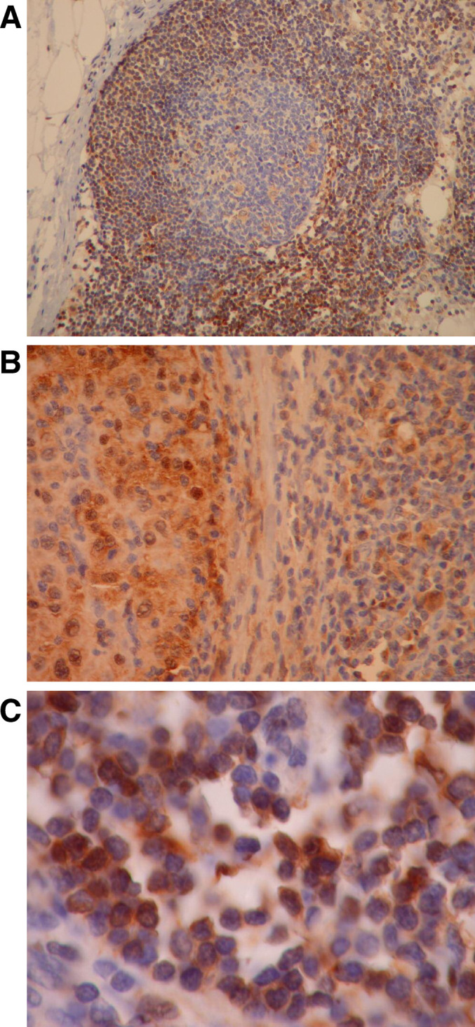
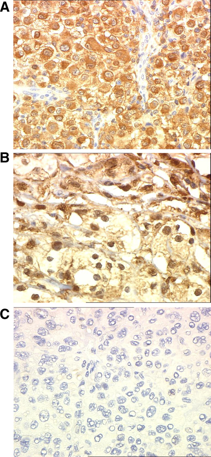
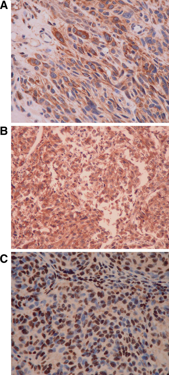
Interferon-alpha (IFN-alpha) is used as an adjuvant therapy in patients with malignant melanoma and who have undergone surgical resection of high-risk lesions. Defective expression or activation of STAT1 or STAT2 has been shown to correlate with IFN-alpha or resistance in vitro; however, recent data from our laboratory suggest that the anti-tumor effects of IFN-alpha are dependent on STAT1 signaling within host immune cells. We measured STAT1 and STAT2 expression in 28 melanoma biopsies (8 cutaneous lesions; 1 lung metastasis; 19 nodal metastases) obtained from patients prior to the initiation of adjuvant IFN-alpha therapy. Disease recurrence following IFN-alpha treatment did not correlate with the staining intensity of either STAT1 (P = 0.61) or STAT2 (P = 0.52). Tumors with minimal STAT1 or STAT2 expression (< 20% positive) were present in four patients with tumor-positive lymph nodes, who exhibited prolonged relapse-free survival (> 44 months) following adjuvant therapy. Conversely, high levels of STAT1 were present in a patient who recurred during the course of IFN-alpha therapy. A case study of one patient who experienced recurrent disease during IFN-alpha treatment revealed that STAT1 levels were greater in the recurrent tumor when compared to the original lesion. These studies provide direct evidence to suggest that levels of STAT1 and STAT2 within the tumor do not influence a patient's response to adjuvant IFN-alpha.
Figures

Similar articles
-
Multiparametric flow cytometric analysis of inter-patient variation in STAT1 phosphorylation following interferon Alfa immunotherapy.J Natl Cancer Inst. 2004 Sep 1;96(17):1331-42. doi: 10.1093/jnci/djh252. J Natl Cancer Inst. 2004. PMID: 15339971
-
Resistance to interferons in melanoma cells does not correlate with the expression or activation of signal transducer and activator of transcription 1 (Stat1).J Interferon Cytokine Res. 2002 May;22(5):603-13. doi: 10.1089/10799900252982089. J Interferon Cytokine Res. 2002. PMID: 12060499
-
STAT5 expression correlates with recurrence and survival in melanoma patients treated with interferon-α.Melanoma Res. 2018 Jun;28(3):204-210. doi: 10.1097/CMR.0000000000000435. Melanoma Res. 2018. PMID: 29485532
-
Adjuvant therapy in melanoma.Onkologie. 2003 Jun;26(3):227-33. doi: 10.1159/000071617. Onkologie. 2003. PMID: 12845206 Review.
-
Recent advances in the care of the patient with malignant melanoma.Ann Surg. 1997 Jan;225(1):1-14. doi: 10.1097/00000658-199701000-00001. Ann Surg. 1997. PMID: 8998115 Free PMC article. Review.
Cited by
-
STAT3 Inhibition Induces Apoptosis in Cancer Cells Independent of STAT1 or STAT2.J Mol Biochem. 2013 Feb 20;2(1):18-26. J Mol Biochem. 2013. PMID: 25364701 Free PMC article.
-
Interferon Alpha Signalling and Its Relevance for the Upregulatory Effect of Transporter Proteins Associated with Antigen Processing (TAP) in Patients with Malignant Melanoma.PLoS One. 2016 Jan 6;11(1):e0146325. doi: 10.1371/journal.pone.0146325. eCollection 2016. PLoS One. 2016. PMID: 26735690 Free PMC article.
-
Molecular profiling of microinvasive breast cancer microenvironment progression.J Transl Med. 2019 Jun 3;17(1):187. doi: 10.1186/s12967-019-1936-x. J Transl Med. 2019. PMID: 31159827 Free PMC article.
-
Development of IFN-gamma resistance is associated with attenuation of SOCS genes induction and constitutive expression of SOCS 3 in melanoma cells.Br J Cancer. 2007 Jul 16;97(2):231-7. doi: 10.1038/sj.bjc.6603849. Epub 2007 Jun 19. Br J Cancer. 2007. PMID: 17579625 Free PMC article.
-
Unphosphorylated STAT1 prolongs the expression of interferon-induced immune regulatory genes.Proc Natl Acad Sci U S A. 2009 Jun 9;106(23):9373-8. doi: 10.1073/pnas.0903487106. Epub 2009 May 28. Proc Natl Acad Sci U S A. 2009. PMID: 19478064 Free PMC article.
References
-
- Carson WE. Interferon-alpha-induced activation of signal transducer and activator of transcription proteins in malignant melanoma. Clin Cancer Res. 1998;4:2219. - PubMed
-
- Creagan ET, Dalton RJ, Ahmann DL, Jung SH, Morton RF, Langdon RM, Jr, Kugler J, Rodrigue LJ. Randomized, surgical adjuvant clinical trial of recombinant interferon alfa-2a in selected patients with malignant melanoma. J Clin Oncol. 1995;13:2776. - PubMed
-
- Chawla-Sarkar M, Leaman DW, Jacobs BS, Tuthill RJ, Chatterjee-Kishore M, Stark GR, Borden EC. Resistance to interferons in melanoma cells does not correlate with the expression or activation of signal transducer and activator of transcription 1 (stat1) J Interferon Cytokine Res. 2002;22:603. doi: 10.1089/10799900252982089. - DOI - PubMed
Publication types
MeSH terms
Substances
Grants and funding
LinkOut - more resources
Full Text Sources
Other Literature Sources
Medical
Research Materials
Miscellaneous
