

Three-dimensional structure of HIV-1 virus-like particles by electron cryotomography
- PMID: 15670606
- PMCID: PMC6608732
- DOI: 10.1016/j.jmb.2004.11.064
Three-dimensional structure of HIV-1 virus-like particles by electron cryotomography
Abstract
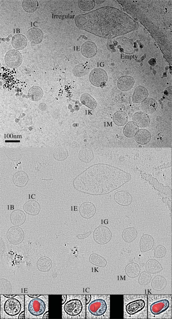
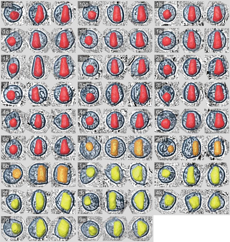
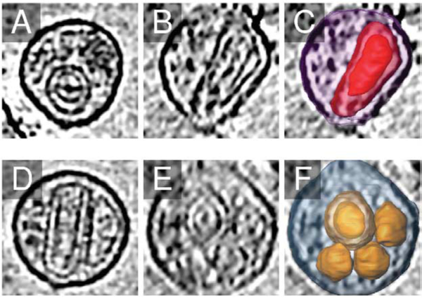
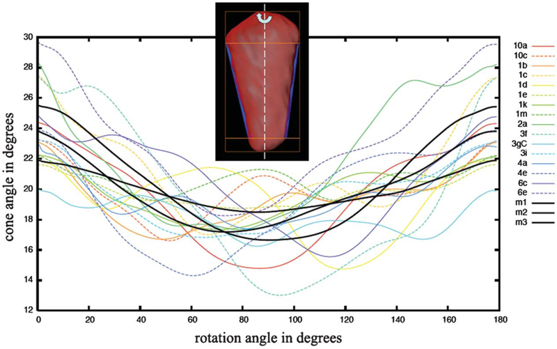
While the structures of nearly every HIV-1 protein are known in atomic detail from X-ray crystallography and NMR spectroscopy, many questions remain about how the individual proteins are arranged in the mature infectious viral particle. Here, we report the three-dimensional structures of individual HIV-1 virus-like particles (VLPs) as obtained by electron cryotomography. These reconstructions revealed that while the structures and positions of the conical cores within each VLP were unique, they exhibited several surprisingly consistent features, including similarities in the size and shape of the wide end of the capsid (the "base"), uniform positioning of the base and other regions of the capsid 11nm away from the envelope/MA layer, a cone angle that typically varied from 24 degrees to 18 degrees around the long axis of the cone, and an internal density (presumably part of the NC/RNA complex) cupped within the base. Multiple and nested capsids were observed. These results support the fullerene cone model for the viral capsid, indicate that viral maturation involves a free re-organization of the capsid shell rather than a continuous condensation, imply that capsid assembly is both concentration-driven and template-driven, suggest that specific interactions exist between the capsid and the adjacent envelope/MA and NC/RNA layers, and show that a particular capsid shape is favored strongly in-vivo.
Figures
References
-
- Gelderblom HR, Hausmann EHS, Ozel M, Pauli G & Koch MA (1987). Fine-structure of human immunodeficiency virus (HIV) and immuno-localization of structural proteins. Micron Microsc. Acta, 18, 335–336. - PubMed
-
- Goto T, Nakai M & Ikuta K (1998). The life-cycle of human immunodeficiency virus type 1. Micron, 29, 123–318. - PubMed
-
- Briggs JAG, Simon MN, Gross I, Krausslich HG, Fuller SD, Vogt VM & Johnson MC (2004). The stoichiometry of Gag protein in HIV-1. Nature Struct. Mol. Biol. 11, 672–675. - PubMed
-
- Hoglund S, Ofverstedt LG, Nilsson A, Lundquist P, Gelderblom H, Ozel M & Skoglund U (1992). Spatial visualization of the maturing HIV-1 core and its linkage to the envelope. AIDS Res. Hum. Retro-viruses, 8,1–7. - PubMed
Publication types
MeSH terms
Substances
Grants and funding
LinkOut - more resources
Full Text Sources
