. 2005 Jan 26:5:5.
doi: 10.1186/1471-2180-5-5.

Surface expression, single-channel analysis and membrane topology of recombinant Chlamydia trachomatis Major Outer Membrane Protein

Affiliations

Surface expression, single-channel analysis and membrane topology of recombinant Chlamydia trachomatis Major Outer Membrane Protein

Heather E Findlay et al. BMC Microbiol. .

Abstract

Background: Chlamydial bacteria are obligate intracellular pathogens containing a cysteine-rich porin (Major Outer Membrane Protein, MOMP) with important structural and, in many species, immunity-related roles. MOMP forms extensive disulphide bonds with other chlamydial proteins, and is difficult to purify. Leaderless, recombinant MOMPs expressed in E. coli have yet to be refolded from inclusion bodies, and although leadered MOMP can be expressed in E. coli cells, it often misfolds and aggregates. We aimed to improve the surface expression of correctly folded MOMP to investigate the membrane topology of the protein, and provide a system to display native and modified MOMP epitopes.

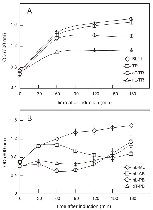
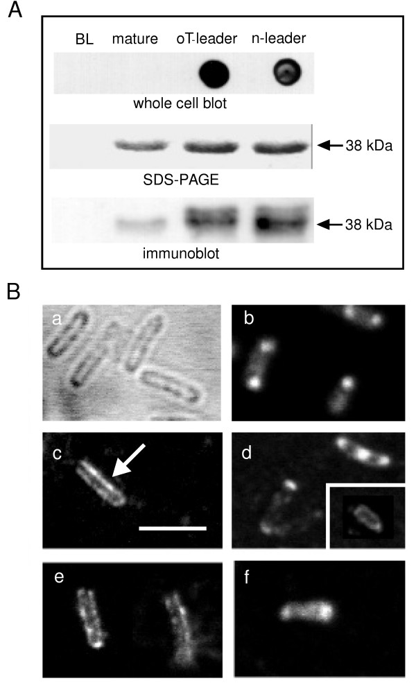
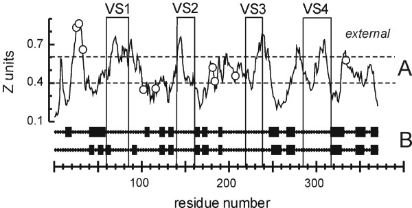
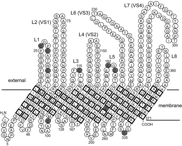
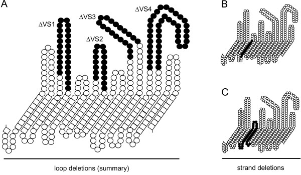
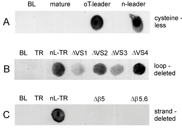
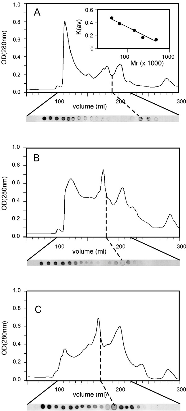
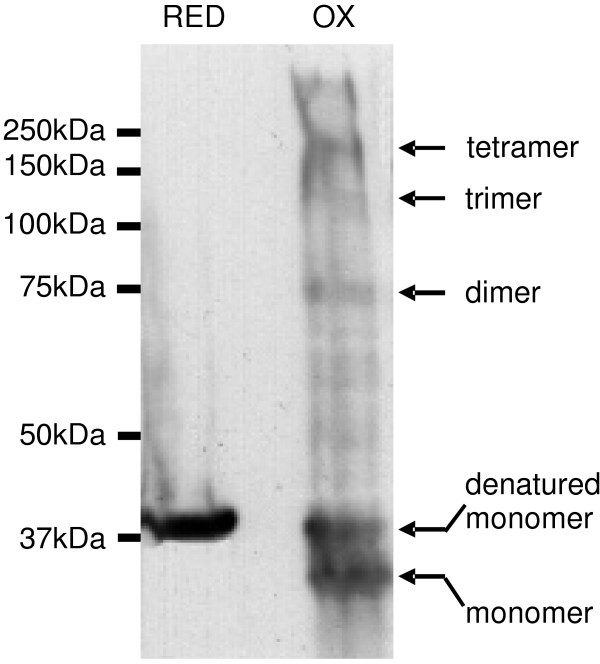
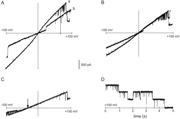
Results: C. trachomatis MOMP was expressed on the surface of E. coli cells (including "porin knockout" cells) after optimizing leader sequence, temperature and medium composition, and the protein was functionally reconstituted at the single-channel level to confirm it was folded correctly. Recombinant MOMP formed oligomers even in the absence of its 9 cysteine residues, and the unmodified protein also formed inter- and intra-subunit disulphide bonds. Its topology was modeled as a (16-stranded) beta-barrel, and specific structural predictions were tested by removing each of the four putative surface-exposed loops corresponding to highly immunogenic variable sequence (VS) domains, and one or two of the putative transmembrane strands. The deletion of predicted external loops did not prevent folding and incorporation of MOMP into the E. coli outer membrane, in contrast to the removal of predicted transmembrane strands.

Conclusions: C. trachomatis MOMP was functionally expressed on the surface of E. coli cells under newly optimized conditions. Tests of its predicted membrane topology were consistent with beta-barrel oligomers in which major immunogenic regions are displayed on surface-exposed loops. Functional surface expression, coupled with improved understanding of MOMP's topology, could provide modified antigens for immunological studies and vaccination, including live subunit vaccines, and might be useful to co-express MOMP with other chlamydial membrane proteins.


Figures

Figure 1
Figure 1
Effects of leader sequences on the growth of E. coli BL21 cells expressing chlamydial porins. A. Moderate inhibition of bacterial (BL21) growth during expression of C. trachomatis MOMP containing native (nL-TR) and OmpT (oT-TR) leaders, contrasting with lack of significant inhibition during expression of mature, leaderless C. trachomatis MOMP (TR). B. Slight inhibition of bacterial growth during expression of C. muridarum MOMP with its native leader (nL-MU, compare to BL21 in A), contrasting with strong inhibition when Ch. abortus MOMP is expressed with its native leader (nL-AB). Cell growth is markedly reduced with both native-leadered PorB (nL-PB) and OmpT-leadered PorB (oT-PB). Note the "recovery" as non-resistant organisms overgrow in β-lactamase-containing cultures (see text). Measurements are means ± SEM (n = 4 independent experiments).
Figure 2
Figure 2
Expression and processing of C. trachomatis MOMP in BL21omp8 cells. Coomassie-stained 12% (w/v) SDS-PAGE of OM proteins extracted using 1%(w/v) OG (10 μg protein per lane) from cells induced for 4 hrs in LB medium, showing dependence of C. trachomatis MOMP expression and processing on temperature and leader sequence. Cells incubated at 16°C and 37°C were induced with 0.1 mM IPTG and 1 mM IPTG, respectively. omp8: non-transformed omp8 cells; oT-TR: cells expressing C. trachomatis MOMP with the OmpT leader; nL-TR: cells expressing C. trachomatis MOMP with its native leader.
Figure 3
Figure 3
Insertion of MOMP into the E. coli outer membrane. A. Recombinant C. trachomatis MOMP was expressed for 2 hrs at 25°C from constructs encoding either no leader (mature), the OmpT (oT) leader, or the native (n) leader, and immunodetected on the surface of intact BL21 cells (upper panel) using a specific anti-MOMP polyclonal Ab. Non-expressing BL21 cells (BL) show no signal, and mature, leaderless MOMP does not reach the cell surface. The middle and lower panels show, respectively, SDS-PAGE and immunoblot analyses of the corresponding recombinant proteins under these conditions (prepared as in Fig. 2). Note the presence of some unprocessed protein, revealed by the immunoblots of leadered protein expression. Representative of 3 similar experiments. B. Immunofluorescence confocal microscopy (panels b–f), with examples of unstained cells (panel a); cells permeabilised and stained after expressing mature, non-leadered MOMP (panel b); cells expressing OmpT-leadered MOMP stained before (panel c) and after permeabilisation (panel d, with inset permeabilised omp8 cell after 12 hrs induction at 16°C); cells expressing native-leadered MOMP under corresponding conditions (panels e and f, respectively). The scale bar (panel c) is 3 μm, and the arrow points out membrane staining. Representative of 3 similar experiments.
Figure 4
Figure 4
Membrane topology and secondary structure predictions for C. trachomatis MOMP. A. "Membrane crossing" prediction. Surface-exposed VS domains and cysteine residues are indicated by boxes and circles, respectively. A "complete membrane crossing" corresponds to a (contiguous) region of the plot that crosses both dotted lines in sequence, where the dotted lines represent the internal (periplasmic) and external borders of the outer membrane (in Z units). B. Two independent β-strand predictions, TMBETA [38] (upper line) and B2TMPRED [37] (below). The strands are boxed. The residue numbers refer to the mature (processed) protein.
Figure 5
Figure 5
Model of C. trachomatis MOMP. The MOMP β-barrel is broken apart at the putative N-C salt bridge in β-strand 16 to display the protein in a 2-D projection. Residues in the transmembrane strands are boxed, with a bold border to indicate side chains facing the bilayer. External loops (including constituent VS domains) are labeled, and cysteine residues are shaded.
Figure 6
Figure 6
Loop and strand deletion maps. A. Summary of individual loop deletions (filled residues) providing four C. trachomatis MOMP proteins deficient in VS domains 1, 2, 3 or 4 (designated ΔVS1-4, respectively). B. Deletion of predicted β-strand 5 and its associated internal loop, designated in the text as Δβ5. C Deletion of predicted β-strands 5 and 6 and their associated internal and external loops, designated in the text as Δβ5,6.
Figure 7
Figure 7
Effects of cysteine mutagenesis and loop and strand deletions on surface exposure of MOMP. A. Replacement of all 9 cysteine residues in C. trachomatis MOMP by alanine residues fails to prevent surface exposure. BL: non-expressing BL21 cells; mature: cells expressing mature MOMP without a leader sequence; oT-leader and n-leader, cells expressing MOMPs with ompT and native leaders, respectively. B. Removal of loops containing putative external VS domains (shown in Fig. 6A) fails to prevent surface exposure. TR: cells expressing mature MOMP; nL-TR: unmodified MOMP with its native leader. C. Removal of putative β-strand regions illustrated in Fig. 6B–C prevents surface exposure. Each result is representative of at least 3 similar experiments.
Figure 8
Figure 8
Gel-exclusion chromatography of solubilised MOMPs. A. GE analysis of C. trachomatis MOMP expressed in BL21omp8 cells with the OmpT leader, solubilised from an OM preparation in 1% (w/v) LDAO. Immuno-dotblots from successive 5 ml fractions between 100–220 ml (shown below the absorbance trace) reveal the appearance of high-molecular mass MOMP aggregates after the void volume, with a second immunoreactive peak at 195 ml (range 185–205 ml), corresponding to a molecular mass of 80 kDa (note inset column calibration trace, also in the presence of detergent). B. GE analysis of C. trachomatis MOMP expressed as in A but solubilised in 1% (w/v) Zwittergent 3–14. Oligomeric MOMP peaks at 180 ml (range 170–190 ml), corresponding to a molecular mass of 120 kDa. C. GE analysis of C. trachomatis MOMP in which all 9 cysteines were changed to alanine, expressed and solubilised as in B. Oligomeric MOMP peaks at 170 ml (range 160–190 ml), corresponding to a molecular mass of 160 kDa. Each trace is representative of at least 3 experiments.
Figure 9
Figure 9
Cysteine cross-linking of recombinant C. trachomatis MOMP. MOMP expressed in BL21omp8 cells with the OmpT leader was covalently cross-linked as described in the text and OM proteins were separated by reducing (RED) and non-reducing (OX) SDS-PAGE, followed by Western blotting using a specific anti-MOMP antibody. The positions of molecular mass markers are shown to the left, and the proposed identities of bands are shown to the right.
Figure 10
Figure 10
Single-channel analysis of recombinant C. trachomatis MOMP from BL21omp8 cells. A. Channel currents in response to two transmembrane voltage ramps (-100 mV to +100 mV), with 1 M KCl cis and trans. Up to 3 channels are open during ramp (a), and 2 of the channels close in succession at about +80 mV. In ramp (b), 1 channel is open at 0 mV, and 2 channels are open at +100 mV. B. Channel currents in the presence of 500 mM KCl cis and trans during two voltage ramps (as in A). Note the reduction in single-current amplitudes, and current reversal at 0 mV as in A. C. Currents during two voltage ramps in asymmetric KCl, 500 mM cis vs 50 mM trans. Note the shift in equilibrium potential (Er) from 0 mV to -30 mV (arrowed). Positive (upgoing) currents at 0 mV indicate a net flux of K+ cis to trans in the absence of an electrical driving force. D. Equilibrium currents in symmetric 500 mM KCl at a constant holding potential of +100 mV, showing brief single-channel closures and voltage-dependent inactivation of all 3 channels within 5 s.

Similar articles

Cited by

References

    1. Stephens ES, ed . Chlamydia intracellular biology, pathogenesis, and immunity. ASM Press, Washington, DC; 1999.
    1. Hatch TP. Disulfide cross-linked envelope proteins: the functional equivalent of peptidoglycan in chlamydiae? J Bacteriol. 1996;178:1–5. - PMC - PubMed
    1. Caldwell HD, Kromhout J, Schachter J. Purification and partial characterization of the major outer membrane protein of Chlamydia trachomatis. Infect Immun. 1981;31:1161–11765. - PMC - PubMed
    1. Salari SH, Ward ME. Polypeptide composition of Chlamydia trachomatis. J Gen Microbiol. 1981;123:197–207. - PubMed
    1. Bavoil P, Ohlin A, Schachter J. Role of disulfide bonding in outer membrane structure and permeability in Chlamydia trachomatis. Infect Immun. 1984;44:479–485. - PMC - PubMed

Publication types

LinkOut - more resources