. 2005 Apr;16(4):1800-10.
doi: 10.1091/mbc.e04-09-0788. Epub 2005 Jan 26.

Identification of xenopus CENP-A and an associated centromeric DNA repeat

Affiliations

Identification of xenopus CENP-A and an associated centromeric DNA repeat

Nathaniel S Edwards et al. Mol Biol Cell. 2005 Apr.

Abstract

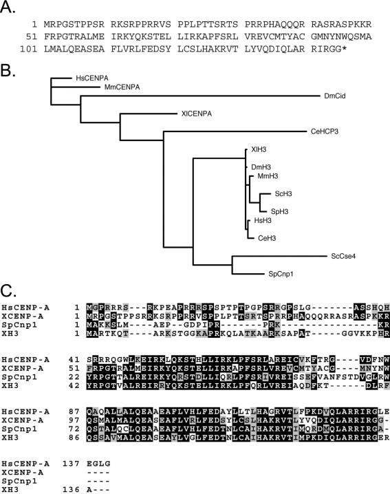
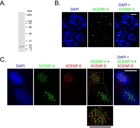
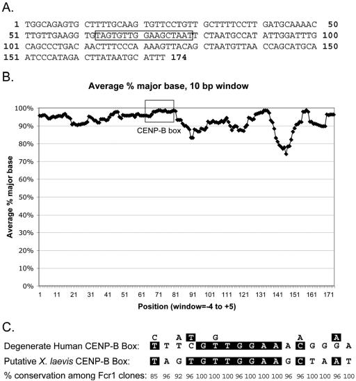
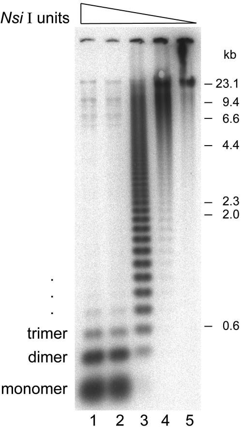
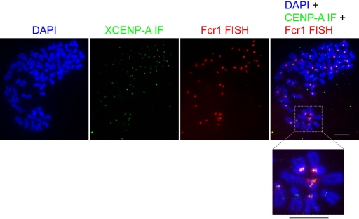
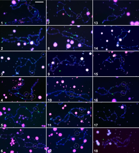
Kinetochores are the proteinaceous complexes that assemble on centromeric DNA and direct eukaryotic chromosome segregation. The mechanisms by which higher eukaryotic cells define centromeres are poorly understood. Possible molecular contributors to centromere specification include the underlying DNA sequences and epigenetic factors such as binding of the centromeric histone centromere protein A (CENP-A). Frog egg extracts are an attractive system for studying centromere definition and kinetochore assembly. To facilitate such studies, we cloned a Xenopus laevis homologue of CENP-A (XCENP-A). We identified centromere-associated DNA sequences by cloning fragments of DNA that copurified with XCENP-A by chromatin immunoprecipitation. XCENP-A associates with frog centromeric repeat 1 (Fcr1), a 174-base pair repeat containing a possible CENP-B box. Southern blots of partially digested genomic DNA revealed large ordered arrays of Fcr1 in the genome. Fluorescent in situ hybridization with Fcr1 probes stained most centromeres in cultured cells. By staining lampbrush chromosomes, we specifically identified the 11 (of 18) chromosomes that stain consistently with Fcr1 probes.


Figures

Figure 1.
Figure 1.
Identification of a sequence homologue of CENP-A in X. laevis. (A) Predicted amino acid sequence of XCENP-A. The asterisk denotes the stop codon. (B) Phylogenetic tree depicting probable evolutionary relationships between CENP-A and H3 homologues from humans (HsCENP-A and HsH3, respectively), mice (MmCENP-A and MmH3), frogs (XCENP-A and XH3), fruit flies (DmCid and DmH3), worms (CeHCP-3 and CeH3), fission yeast (SpCnp1 and SpH3), and budding yeast (ScCse4 and ScH3). Branch lengths are proportional to predicted evolutionary distances. The tree was generated with MrBayes (Ronquist and Huelsenbeck, 2003). (C) Multiple sequence alignment of CENP-A homologues from fission yeast, humans, and frogs. The X. laevis H3 protein sequence is included in the alignment for reference. Dark shading indicates identical residues; light shading indicates similar residues.
Figure 2.
Figure 2.
XCENP-A localizes to kinetochores. (A) Anti-XCENP-A Western blot of nuclear proteins reveals a single prominent band with mobility consistent with the predicted molecular mass of XCENP-A (17 kDa). (B) Indirect immunofluorescence against XCENP-A (green) on DAPI-stained metaphase chromosome spreads (blue) from cultured cells. (C) Direct immunofluorescence on mitotic (top left) and interphase (bottom right) cells. Cells were stained with directly labeled antibodies against XCENP-A (green) and XCENP-E (red). DAPI-stained DNA is shown in blue. Boxed region is enlarged below. Bars, 10 μm. XCENP-A and XCENP-E foci seem to be adjacent rather than identical. This pattern might reflect the expected juxtaposition of inner and outer kinetochore components or the two images not being properly in register with each other.
Figure 3.
Figure 3.
Identification of Fcr1, a 174-base pair centromere-associated repeat. (A) Consensus sequence of the monomer unit (with ends defined arbitrarily) of the DNA satellite cloned from α-CENP-A chromatin immunoprecipitates. The box indicates the putative CENP-B box (positions 63 through 79). (B) Conservation of Fcr1 sequences among 38 analyzed monomeric units and submonomeric fragments. The graph plots average (over a 10-base pair window) percentage of sequences containing the consensus nucleotide at each position. Boxed region indicates putative CENP-B box. (C) Similarity between a 17-base pair element in Fcr1 (“putative X. laevis CENP-B box”) and the degenerate human CENP-B box (Choo, 1997). The percentage of Fcr1 clones containing the consensus nucleotide is shown below each position.
Figure 4.
Figure 4.
Fcr1 is present in large ordered arrays in the frog genome. Southern blot of Nsi I-digested genomic DNA with radioactive probe for Fcr1. Genomic DNA (1 μg per lane) was digested with 25 U (lane 1), 5 U (lane 2), 1 U (lane 3), 0.2 U (lane 4), or 0.04 U (lane 5) of Nsi I.
Figure 5.
Figure 5.
In situ hybridization against Fcr1 stains most centromeres. Fluorescent in situ hybridization of digoxigenin-labeled Fcr1 probe (red) against DAPI-stained metaphase chromosome spreads (blue) from cultured cells. Indirect immunofluorescence against XCENP-A is shown in green. Boxed region is enlarged below. Bars, 10 μm.
Figure 6.
Figure 6.
Fcr1 hybridization against lampbrush chromosomes confirms consistent staining of 11 of the 18 frog chromosomes. Each of the 18 X. laevis lampbrush chromosomes (stained with DAPI, blue) can be identified by the distinct patterns of loci stained by antibodies against RNA polymerase III (green) (Murphy et al., 2002). Fluorescent in situ hybridization with Cy3-labeled Fcr1 probe is shown in red. Chromosomes are numbered in descending order according to length (Murphy et al., 2002). This composite combines representative images from an average of three analyzed examples of each of the 18 lampbrush chromosomes. Cross-hybridization with extrachromosomal nucleoli (large DAPI- and Fcr1-stained spheres) is believed to be caused by polylinker sequences at the ends of the Fcr1 probe, which can hybridize with rRNA (Witkiewicz et al., 1993). Dashed circles highlight chromosomal sites of Fcr1 hybridization. Bar, 20 μm.

References

    1. Alonso, A., Mahmood, R., Li, S., Cheung, F., Yoda, K., and Warburton, P. E. (2003). Genomic microarray analysis reveals distinct locations for the CENP-A binding domains in three human chromosome 13q32 neocentromeres. Hum. Mol. Genet. 12, 2711–2721. - PubMed
    1. Amor, D. J., Bentley, K., Ryan, J., Perry, J., Wong, L., Slater, H., and Choo, K. H. (2004). Human centromere repositioning “in progress.” Proc. Natl. Acad. Sci. USA 101, 6542–6547. - PMC - PubMed
    1. Amor, D. J., and Choo, K. H. (2002). Neocentromeres: role in human disease, evolution, and centromere study. Am. J. Hum. Genet. 71, 695–714. - PMC - PubMed
    1. Ando, S., Yang, H., Nozaki, N., Okazaki, T., and Yoda, K. (2002). CENP-A, -B, and -C chromatin complex that contains the I-type alpha-satellite array constitutes the prekinetochore in HeLa cells. Mol. Cell. Biol. 22, 2229–2241. - PMC - PubMed
    1. Biggins, S., and Walczak, C. E. (2003). Captivating capture: how microtubules attach to kinetochores. Curr. Biol. 13, R449–R460. - PubMed

Publication types

MeSH terms

LinkOut - more resources