Comparative Study
. 2005 Jan 26;25(4):778-91.
doi: 10.1523/JNEUROSCI.4235-04.2005.

Differential transport and local translation of cytoskeletal, injury-response, and neurodegeneration protein mRNAs in axons

Affiliations
Comparative Study

Differential transport and local translation of cytoskeletal, injury-response, and neurodegeneration protein mRNAs in axons

Dianna Willis et al. J Neurosci. .

Erratum in

  • J Neurosci. 2010 Nov 10;30(45):np. Smit, August [corrected to Smit, August B]

Abstract

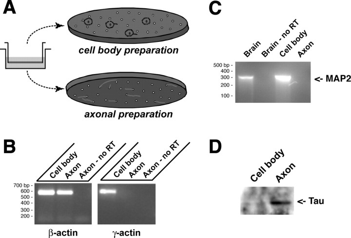
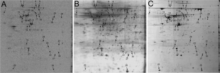
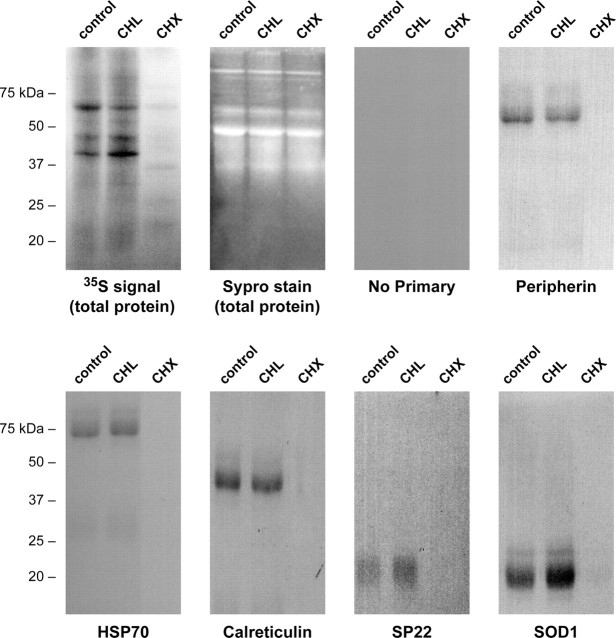
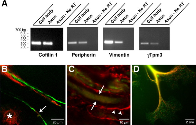
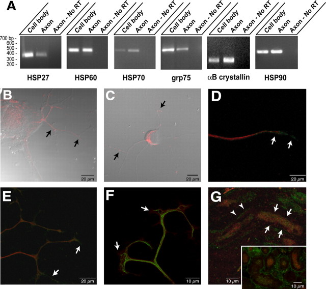
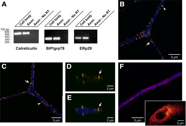
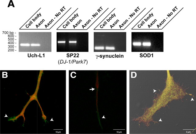
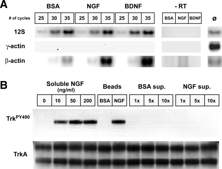
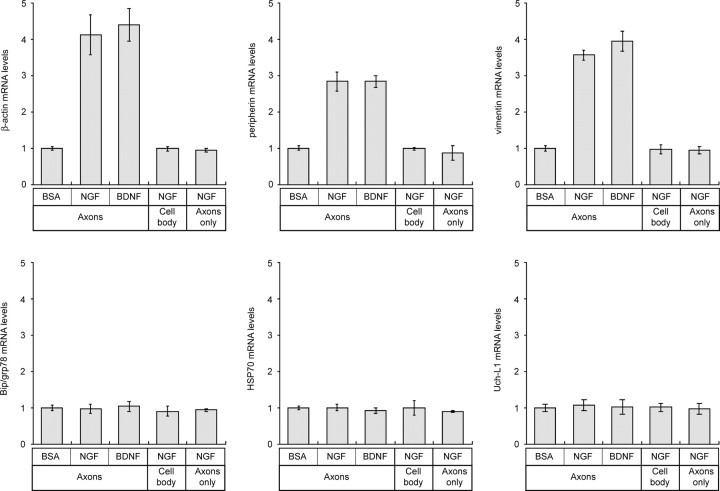
Recent studies have begun to focus on the signals that regulate axonal protein synthesis and the functional significance of localized protein synthesis. However, identification of proteins that are synthesized in mammalian axons has been mainly based on predictions. Here, we used axons purified from cultures of injury-conditioned adult dorsal root ganglion (DRG) neurons and proteomics methodology to identify axonally synthesized proteins. Reverse transcription (RT)-PCR from axonal preparations was used to confirm that the mRNA for each identified protein extended into the DRG axons. Proteins and the encoding mRNAs for the cytoskeletal proteins beta-actin, peripherin, vimentin, gamma-tropomyosin 3, and cofilin 1 were present in the axonal preparations. In addition to the cytoskeletal elements, several heat shock proteins (HSP27, HSP60, HSP70, grp75, alphaB crystallin), resident endoplasmic reticulum (ER) proteins (calreticulin, grp78/BiP, ERp29), proteins associated with neurodegenerative diseases (ubiquitin C-terminal hydrolase L1, rat ortholog of human DJ-1/Park7, gamma-synuclein, superoxide dismutase 1), anti-oxidant proteins (peroxiredoxins 1 and 6), and metabolic proteins (e.g., phosphoglycerate kinase 1 (PGK 1), alpha enolase, aldolase C/Zebrin II) were included among the axonally synthesized proteins. Detection of the mRNAs encoding each of the axonally synthesized proteins identified by mass spectrometry in the axonal compartment indicates that the DRG axons have the potential to synthesize a complex population of proteins. Local treatment of the DRG axons with NGF or BDNF increased levels of cytoskeletal mRNAs into the axonal compartment by twofold to fivefold but had no effect on levels of the other axonal mRNAs studied. Neurotrophins selectively increased transport of beta-actin, peripherin, and vimentin mRNAs from the cell body into the axons rather than changing transcription or mRNA survival in the axonal compartment.


Figures

Figure 1.
Figure 1.
Isolation of DRG axons. A, Schematic of the DRG culture method for isolation of axons is illustrated. Dissociated DRGs are cultured in tissue culture insert with polyethylene tetraphthalate membrane with 8-μm-diameter pores shown to the left. The neuron cell bodies (green) and non-neuronal cells (red) remain on the upper membrane surface. Axons traverse to the underside of the membrane through the pores. B, β-Actin was amplified using RNA from either the cell body or the axons as a template, but γ-actin mRNA could only be amplified from the cell-body RNA. Axonal RNA processed for RT-PCR without the addition of reverse transcriptase shows that the β-actin PCR product detected in the axons is specific for amplification of mRNA. C, RT-PCR was used to determine whether axons contained MAP2 mRNA. In this case, rat brain RNA was used for both positive and “no RT” control for PCR. Note that the DRG cell body contains even more MAP2 mRNA than does the total rat brain RNA, but no MAP2 was amplified from the axonal preparation. D, Protein extracted from the cell body and axonal preparations (10 μg/lane) was separated by 10% SDS-PAGE and transferred to PVDF membrane. By immunoblotting, a band corresponding to tau was clearly visible in the axonal preparation, but only a faint band can be seen in the cell-body fraction.
Figure 2.
Figure 2.
Two-dimensional separation of radiolabeled isolated axons. Representative images from 2D gels (first dimension, pH 4-7; second dimension, 11% SDS-PAGE) from 35S-labeled axonal preparations (A, B) and preparative gel from whole DRG culture lysates (C) are shown. Approximately the 4 × 10⁶ cpm was fractionated by 2D gel electrophoresis in the gel shown in A and B. A shows the image of radioactive proteins by phosphorimaging; B shows the aurodye stain for total proteins in the blot from A. C shows preparative gel of whole DRG lysates that was stained with Coomassie. The numbered spots indicate proteins that could be aligned between the gels. These spots were excised from the preparative gel and processed for MS analyses (Table 2). Images of pH 3-10 gels are included in supplemental data (Fig. S1, available at www.jneurosci.org as supplemental material).
Figure 3.
Figure 3.
Axoplasmic protein synthesis. Sheared axons were pretreated with 25 μg/ml chloramphenicol (CHL), 10 μg/ml cycloheximide (CHX), or vehicle (Control) for 20 min, and then 2 mCi/ml [35S]methionine/cysteine was added for 4 h. The first two panels show fractionated lysates from the axons with total protein visualized by Sypro Ruby stain (Sypro) and labeled proteins visualized by autoradiography (3 d exposure). The remainder of the panels show autoradiograms of immunoprecipitated proteins indicated (peripherin, ≈54 kDa; HSP70, ≈70 kDa; calreticulin, ≈48 kDa; Uch-L1, ≈25 kDa; SP22, ≈20 kDa; SOD1, ≈15 kDa). Note that newly synthesized proteins are detected in the control and CHL lanes but not in the CHX lane, whereas the Sypro stain of the lysates before immunoprecipitation shows approximately equivalent levels of unlabeled proteins in all lanes.
Figure 4.
Figure 4.
Cytoskeletal proteins and encoding mRNAs in DRG axons. A, RNA was isolated from axonal and cell-body preparations and processed for RT-PCR as in Figure 1 B. Selective amplification of β-actin mRNA but not γ-actin mRNAs from the axonal RNA confirmed the purity of the axonal preparation (data not shown). Transcript-specific primers confirmed that peripherin, vimentin, γ-Tpm3, and cofilin 1 mRNAs extended into the DRG axons. B, Cultures of injury-conditioned DRG neurons were colabeled with antibodies to peripherin (green) and vimentin (red) and imaged by confocal microscopy. A single optical plane is shown. Note that intra-axonal vimentin and peripherin signals (arrows) are clearly distinct from the vimentin immunoreactive in the Schwann cells (asterisk). C, Sections of crushed sciatic nerve were colabeled for vimentin (red) and peripherin (green). The image displays three-dimensional projection of 10 XY planes taken at 0.25 μm intervals. The arrows indicate an axon where XY planes were only taken directly through the axon, specifically excluding any myelin sheath above or below the axon. Note that vimentin is present both in the axon (arrows) and in Schwann cells of the surrounding myelin sheath (arrowheads). D, Injury-conditioned DRG cultures were colabeled for tropomyosin (green) and neurofilament (red). An epifluorescent image of a large growth cone with tropomyosin and neurofilament signals merged is shown. Note that tropomyosin immunoreactivity extends well into the growth cone.
Figure 5.
Figure 5.
Heat shock and heat shock-like proteins and encoding mRNAs in regenerating axons. A, RT-PCR was performed on axonal versus cell-body RNA isolates using primers specific for αB crystallin, HSP27, HSP60, grp75, HSP70, and HSP90 mRNAs. Purity of the axonal preparation was determined by RT-PCR for β- and γ-actin mRNAs as shown in Figure 1 B. Note that each of these mRNAs was detected in the axonal RNA templates, but the relative amounts vary. HSP27 and grp75 are more abundant in the cell body than in the axon. Approximately equivalent amounts of HSP60 and HSP90 mRNAs are amplified from the cell body and axons, whereas αB crystallin HSP70 mRNA is relatively more abundant in the axons than in the cell-body isolates compared with the other mRNAs examined. B, Injury-conditioned DRG cultures were labeled for HSP60. This representative image displays are constructed three-dimensional projection of HSP60 (red; 15 optical XY planes taken at 0.2 μm intervals) merged with a single differential interference contrast (DIC) image. Arrows show HSP60 immunoreactivity in proximal portions of the DRG axons. C, DRG cultures stained for grp75 signal (red) show intra-axonal grp75 immunoreactivity along the shaft of the axons (arrows) in this three-dimensional projection of grp75 signal (12 optical XY planes taken at 0.3 μm intervals) merged with a single DIC image. D, A single optical XY plane of distal axon from DRG cultures colabeled for HSP70 (green) and peripherin (red). Arrows indicate HSP70 immunoreactivity that extends beyond the peripherin immunoreactivity into the growth cone. E, Colabeling cultures of injury-conditioned DRG neurons for HSP90 (green) and neurofilament (red) shows that HSP90 extends into the distal axon (arrows), similar to HSP70 shown in D, in this three-dimensional projection (12 optical XY planes taken at 0.2 μm intervals). F, A reconstructed three-dimensional projection shows terminal axons of cultures stained for αB crystall in (red) and neurofilament (green) (projection is from 10 optical XY planes taken at ∼0.15 μmintervals). Note that similar to HSP70 and HSP90, αB crystallin immunoreactivity extends into the growth cone beyond the neurofilament signal (arrows). G, Section of sciatic nerve as in F was colabeled for HSP70 (green) and peripherin (red). The full panel shows a longitudinal section where intra-axonal HSP70 signal is visible in an optically isolated axon (arrows) in this projected three-dimensional image. HSP70 signal separate from the peripherin immunoreactivity is also seen in Schwann cell cytoplasm of the myelin sheath adjacent to this axon (arrow-heads). The inset shows a cross section of nerve where intra-axonal HSP70 signal is clearly discerned from the bright signal in the surrounding Schwann cells.
Figure 6.
Figure 6.
Resident-ER proteins and encoding mRNAs in DRG axons. A, RT-PCR for rat grp78/BiP, calreticulin, and ERp29 mRNAs shows that these transcripts extend into the DRG axons. Amplification of β- and γ-actin mRNAs confirmed the absence of any γ-actin mRNA in the axonal sample. B, A single optical XY plane through the axon of a cultured DRG neuron showing calreticulin (green), SERCA (red), and neurofilament (blue) signals. The majority of calreticulin colocalizes with SERCA (arrow) with only occasional calreticulin signal seen in the absence of adjacent SERCA (arrowhead). C, A representative single optical XY plane through the axon of cultured DRG neurons stained for grp78/BiP (green), SERCA (red), and neurofilament (blue). Similar to calreticulin in B, the majority of grp78/BiP signal colocalizes with SERCA (arrow) and is rarely seen directly adjacent to SERCA-immunoreactive granules (arrowhead). D, E, A single XZ plane through the axon illustrated in C taken at the bifurcation level. Signals for grp78/BiP (green) merged with SERCA (red) are shown in D, and grp78/Bip (green), Serca (red), and neurofilament (blue) are merged in E. Note that the grp78/BiP and SERCA immunoreactivity colocalize (arrows) and concentrate along the periphery of the axoplasm. F, A reconstructed three-dimensional confocal image of cultures stained for ERp29 and neurofilament is shown (14 optical XY planes taken at ∼0.12 μm intervals). The panel shows a segment of distal axon with ERp29 in red and neurofilament in blue. The inset shows an adjacent Schwann cell with ERp29 channel intensity displayed as a spectrum, with yellow being the most intense. Similar to signals for calreticulin and grp78/BiP above, ERp29 immunoreactivity appears coarsely granular in the axon and is concentrated along the more peripheral extents of the neurofilament signal. The perinuclear concentration of ERp29 in the Schwann cells is characteristic of ER proteins.
Figure 7.
Figure 7.
Neurodegeneration-associated proteins and encoding mRNAs extend into DRG axons. A, RNA isolated from cell-body versus axonal compartment was processed for RT-PCR using primers specific for Uch-L1, SP22, γ-synuclein, and SOD1 mRNAs. Each of these transcripts extends into the cultured DRG axons. Amplification of β- and γ-actin mRNAs confirmed the purity of the axonal RNA samples (data not shown). B, Image displays a three-dimensional projection through the distal axon of cultured DRG neurons that was reconstructed from 10 XY optical planes taken over 3.5 μm in the z-axis. Note that Uch-L1 immunoreactivity (green) extends beyond the neurofilament signal (red) into the growth cones of this branched axon (arrowheads). C, Image displays a three-dimensional projection through the distal axon of cultured DRG neurons stained for neurofilament (green) and γ-synuclein (red). Note that similar to Uch-L1, γ-synuclein also extends into the distal axon beyond the neurofilament (arrow). A prominent signal for γ-synuclein is also seen along the more proximal segments of the axon shaft (arrowhead). D, Image displays a three-dimensional projection of a growth cone from DRG cultures stained for SP22 (DJ-1/Park7) and SOD1. The SP22 (green) and SOD1 (red) signals represent a reconstruction of seven optical XY planes taken at ∼0.13 μm intervals merged with the DIC image. Note that SP22 and SOD1 extend from axon shaft (arrow) into the growth cone and even into some of the filopodia (arrowheads).
Figure 8.
Figure 8.
Local treatment with neurotrophins regulates axonal mRNA localization. A, The axonal compartments of DRG neuron cultures were selectively treated with NGF-, BDNF-, or BSA-absorbed microparticles for 4 h in the presence of the RNA synthesis inhibitor. The diameter of the microparticles, at more than twice that of the membrane pores, restricted neurotrophin exposure to the axonal compartment. Axonal RNA was normalized to axonal protein content and processed for hot RT-PCR (see Materials and Methods). Total RNA isolated from rat brain was used as a positive control, and reactions performed without addition of RT served as a negative control. Signals for the 12S mitochondrial rRNA showed equivalent levels between the different axonal RNA samples. Absence of any signal for γ-actin mRNA in the axonal samples, even with increased sensitivity of radioactive PCR, indicates pure preparations of axonal RNA. Consistently more β-actin mRNA signal was amplified from the NGF- and BDNF-treated axonal RNA samples compared with the BSA-treated axonal samples. B, To determine whether significant levels of NGF were leached from the beads during the 4 h incubations shown in A, PC12 cells were treated with soluble NGF (0, 10, 50, and 200 ng/ml), BSA- or NGF-absorbed beads, and supernatant (sup.) from BSA- or NGF-absorbed beads for 5 min. The supernatant was used at onefold (1x), fivefold (5x), and 10-fold (10x) volume equivalents to the beads. The top panel shows activation of TrkA detected by immunoblotting with anti-phosphoTrk antibody (TrkPY490) (Chang et al., 2003). The bottom panel shows that total levels of Trk are relatively equivalent in all samples. Note that both soluble and bead-absorbed NGF activated TrkA, but the BSA-absorbed beads and supernatant from BSA- and NGF-absorbed beads showed no detectable phospho-TrkA.
Figure 9.
Figure 9.
Neurotrophins regulate axonal localization of cytoskeletal protein mRNAs. Real-time RT-PCR was used to quantitate levels of specific mRNAs in the axonal RNA samples. Results for β-actin, peripherin, vimentin, grp78/BiP, HSP70, and Uch-L1 are illustrated. grp78/BiP, HSP70, and Uch-L1 signals are representative of transcripts that showed no change in levels after NGF or BDNF treatment (see supplemental Fig. S2, available at www.jneurosci.org as supplemental material). All values are displayed relative to a BSA-treated sample as a control. The top three panels represent cultures in which the axons were treated with microparticles, and axonal RNA was quantitated as in A. The bottom left panel represents values for cultures where axons were treated with microparticles as above, but the cell-body compartment was quantitated to evaluate potential changes in gene expression after neurotrophin treatment. The bottom center panel represents axonal RNA values in which axons isolated from the cell body were treated with microparticles and then RNAs were quantitated to evaluate potential neurotrophin-dependent changes in mRNA survival.

Similar articles

Cited by

References

    1. Akbar M, Lundberg A, Liu K, Vidyadaran S, Wells K, Dolatshad H, Wynn S, Wells D, Latchman D, de Belleroche J (2003) The neuroprotective effects of heat shock protein 27 overexpression in transgenic animals against kainate-induced seizures and hippocampal cell death. J Biol Chem 278: 19956-19965. - PubMed
    1. Aronov S, Aranda G, Behar L, Ginzburg I (2001) Axonal tau mRNA localization coincides with tau protein in living neuronal cells and depends on axonal targeting signal. J Neurosci 21: 6577-6587. - PMC - PubMed
    1. Bamburg JR (1999) Proteins of the ADF/cofilin family: essential regulators of actin dynamics. Annu Rev Cell Dev Biol 15: 185-230. - PubMed
    1. Bamburg JR, Bray D (1987) Distribution and cellular localization of actin depolymerizing factor. J Cell Biol 105: 2817-2825. - PMC - PubMed
    1. Bassell GJ, Zhang H, Byrd AL, Femino AM, Singer RH, Taneja KL, Lifshitz LM, Herman IM, Kosik KS (1998) Sorting of beta-actin mRNA and protein to neurites and growth cones in culture. J Neurosci 18: 251-265. - PMC - PubMed

Publication types

MeSH terms

LinkOut - more resources