. 2005 Jan 26;25(4):808-22.
doi: 10.1523/JNEUROSCI.4464-04.2005.

The Nogo-66 receptor homolog NgR2 is a sialic acid-dependent receptor selective for myelin-associated glycoprotein

Affiliations

The Nogo-66 receptor homolog NgR2 is a sialic acid-dependent receptor selective for myelin-associated glycoprotein

Karthik Venkatesh et al. J Neurosci. .

Abstract

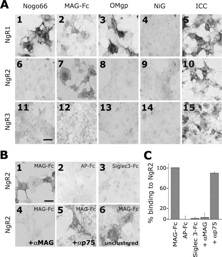
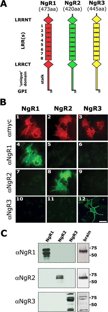
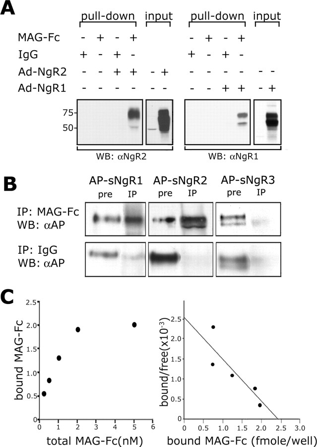
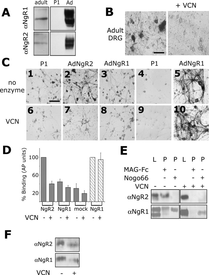
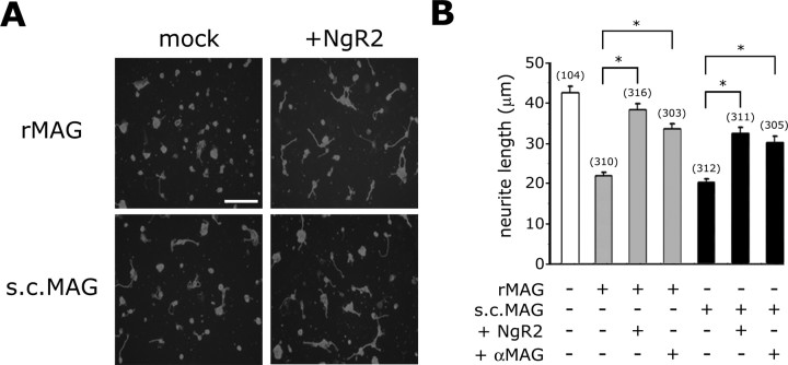
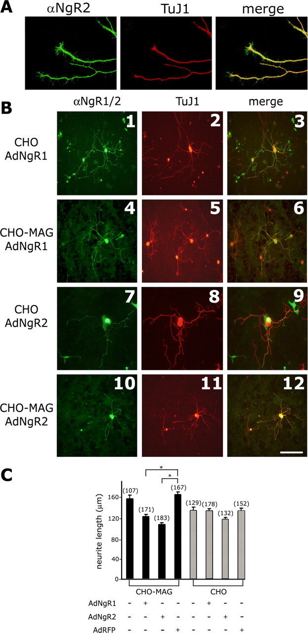
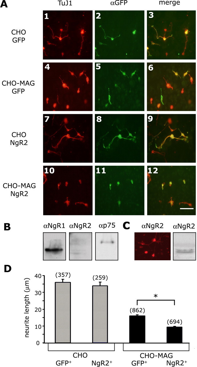
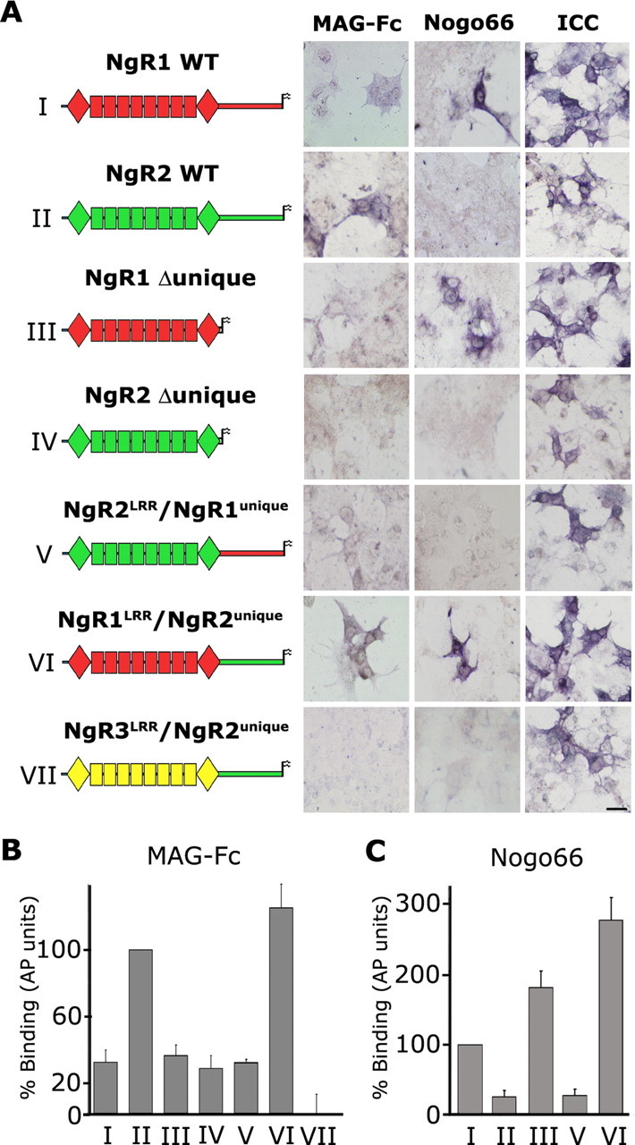
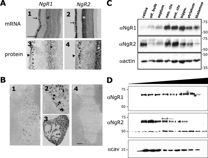
The Nogo-66 receptor (NgR1) is a promiscuous receptor for the myelin inhibitory proteins Nogo/Nogo-66, myelin-associated glycoprotein (MAG), and oligodendrocyte myelin glycoprotein (OMgp). NgR1, an axonal glycoprotein, is the founding member of a protein family composed of the structurally related molecules NgR1, NgR2, and NgR3. Here we show that NgR2 is a novel receptor for MAG and acts selectively to mediate MAG inhibitory responses. MAG binds NgR2 directly and with greater affinity than NgR1. In neurons NgR1 and NgR2 support MAG binding in a sialic acid-dependent Vibrio cholerae neuraminidase-sensitive manner. Forced expression of NgR2 is sufficient to impart MAG inhibition to neonatal sensory neurons. Soluble NgR2 has MAG antagonistic capacity and promotes neuronal growth on MAG and CNS myelin substrate in vitro. Structural studies have revealed that the NgR2 leucine-rich repeat cluster and the NgR2 "unique" domain are necessary for high-affinity MAG binding. Consistent with its role as a neuronal MAG receptor, NgR2 is an axonassociated glycoprotein. In postnatal brain NgR1 and NgR2 are strongly enriched in Triton X-100-insoluble lipid rafts. Neural expression studies of NgR1 and NgR2 have revealed broad and overlapping, yet distinct, distribution in the mature CNS. Taken together, our studies identify NgRs as a family of receptors (or components of receptors) for myelin inhibitors and provide insights into how interactions between MAG and members of the Nogo receptor family function to coordinate myelin inhibitory responses.


Figures

Figure 2.
Figure 2.
Binding profile of myelin inhibitors to Nogo receptors. A, Binding of AP-Nogo-66 (A1, A6, A11), MAG-Fc (A2, A7, A12), OMgp-AP (A3, A8, A13), and AP-NiG (A4, A9, A14) to recombinant NgR1, NgR2, and NgR3 transiently expressed in COS-7 cells. Bound MAG-Fc was detected with anti-human Fc conjugated to AP. Immunocytochemistry with anti-NgR1 (A5), anti-NgR2 (A10), and anti-NgR3 (A15) was used to confirm expression of transfected receptor constructs. B, NgR2 supports binding of oligomerized MAG-Fc (B1), but not AP-Fc (B2) or Siglec 3-Fc (B3). Binding of oligomerized MAG-Fc to NgR2 is blocked by anti-MAG 513 (B4), but not by anti-p75 192 IgG (B5). MAG-Fc that has not been oligomerized with anti-human Fc (unclustered) still binds to NgR2 (B6). C, Quantification of ligand binding in relative AP units. Binding of MAG to NgR2 is equal to 100%. NgR2 does not support AP-Fc or Siglec 3-Fc binding. MAG binding is blocked in the presence of anti-MAG (10 μg/ml), but not anti-p75 (10 μg/ml) IgG. Scale bars: A, B, 20 μm.
Figure 1.
Figure 1.
Characterization of antibodies raised against Nogo receptor family members. A, Domain alignment of the Nogo receptor family members NgR1 (473 amino acid residues), NgR2 (420 amino acid residues), and NgR3 (445 amino acid residues). Nogo receptors are composed of eight canonical leucine-rich repeats (LRR1-LRR8) flanked by cysteine-rich LRRNT and LRRCT subdomains. The highly conserved LRRNT+LRR+LRRCT domains are connected via a more variable unique domain (stalk) to a GPI anchor for membrane attachment. We raised polyclonal antibodies against the distal part of the LRRCT and the unique domain of each of the three NgR family members. B, Immunocytochemistry (ICC) under nonpermeabilizing conditions of myc-tagged NgR1, NgR2, and NgR3 in transfected COS-7 cells reveals abundant cell surface localization of NgR1 (B1), NgR2 (B2), and NgR3 (B3). Anti-NgR1 (B4), anti-NgR2 (B8), and anti-NgR3 (B12) immune sera strongly and selectively react with their cognate antigens. No cross-reactivity with other NgR family members is observed. C, Western blotting with anti-NgR1, anti-NgR2, and anti-NgR3 immune sera allows for selective detection of recombinant (COS-7) and endogenously (adult rat brain) expressed receptors. All three NgR family members are expressed abundantly in the adult brain. Of note, endogenously expressed NgR1 and NgR2 are detected as single bands and migrate at an apparent molecular weight of 65 kDa. Recombinant NgR1 and NgR2 occur as multiple variants between 55 and 70 kDa (Walmsley et al., 2004). Multiple variants are found for recombinant and endogenously expressed NgR3. Scale bar, 20 μm.
Figure 3.
Figure 3.
NgR2 binds MAG directly and with high affinity. A, MAG-Fc affinity precipitation of NgR1 and NgR2 from lysates of Ad-NgR1-transduced (right) or Ad-NgR2-transduced (left) COS-7 cells. Lysates of virus-infected or control (uninfected) cells were subjected to affinity precipitation with MAG-Fc or control IgG. Immunoblotting with anti-NgR1 and anti-NgR2 revealed binding of MAG to NgR2 and NgR1 (pull-down). Of note, MAG binds selectively to the high-molecular-weight forms of NgR2 and preferentially to the higher-molecular-weight forms of NgR1. The input lanes show immunoblots of total cell lysate of control, Ad-NgR1-, and Ad-NgR2-transduced cells. B, MAG-Fc directly binds to soluble NgR1 (AP-sNgR1) and NgR2 (AP-sNgR2), but not to NgR3 (AP-sNgR3). A control IgG does not bind to any of the soluble Nogo receptors. Input (pre) and precipitates (IP) were analyzed by immunoblotting with anti-AP. C, Scatchard analysis of NgR2-transfected COS-7 cells to increasing concentrations of MAG-Fc (0.1-5 nm) produced a linear plot revealing an apparent KD of 2 nm (a representative plot of 3 independent experiments is shown on the right). The graph on the left shows the MAG-Fc saturation binding curve to NgR2-expressing COS-7 cells.
Figure 4.
Figure 4.
Ectopic expression of NgR2 in neurons is sufficient to confer sialic acid-dependent MAG binding. A, Immunoblotting of dissociated adult rat DRG cultures reveals expression of NgR1 and NgR2. Neonatal (P1) DRG neurons express very low levels of NgR1 and NgR2. When transduced with Ad-NgR1 or Ad-NgR2, P1 DRG neurons express high levels of NgR1 and NgR2 as revealed by Western blot analysis. B, Dissociated adult rat DRGs support binding of MAG-Fc in a sialic acid-dependent, VCN-sensitive manner. C, P1-P2 DRGs support MAG-Fc binding weakly (C1). After transduction with Ad-NgR2 (C2) or Ad-NgR1 (C3), the DRGs support MAG-Fc binding more strongly. Binding of MAG-Fc to control (C6), Ad-NgR2-transduced (C7), and Ad-NgR1-transduced (C8) DRGs is sensitive to VCN treatment. Binding of Nogo-66 to Ad-NgR1-transduced DRGs is very robust (C5) compared with control (uninfected) DRGs (C4), and Nogo-66 binding to NgR1 is not sensitive to VCN treatment (C10). D, Quantification of MAG-Fc binding to Ad-NgR2, Ad-NgR1, and mock-transduced DRGs in the absence (-) or presence (+) of VCN and binding of AP-Nogo-66 to Ad-NgR1-transfected DRGs (striped bars). MAG binding to Ad-NgR2 and AP-Nogo-66 binding to Ad-NgR1-transfected cultures is normalized to 100%. Error bars indicate SEM. E, MAG-Fc affinity precipitation of NgR2 and NgR1 from virally transduced DRG cultures is robust and highly sensitive to VCN treatment. Cell lysates (L) show high- and low-molecular-weight forms of NgR1 and NgR2. Western blot analysis of precipitates (P) reveals that MAG preferentially complexes with the higher-molecular-weight forms of NgR1 and NgR2. After VCN treatment NgR1 and NgR2 no longer support the binding of MAG-Fc. Moreover, VCN treatment results in a ∼2-3 kDa decrease in molecular weight of NgR1 and NgR2. Binding of Nogo-66 to Ad-NgR1-transduced DRGs is not sensitive to VCN treatment. F, A similar VCN-dependent drop in molecular weight was observed with endogenously expressed NgR1 and NgR2 isolated from P14 rat brain. Scale bars: B, C, 100 μm.
Figure 5.
Figure 5.
Exogenous NgR2 antagonizes MAG inhibition. A, Neurite outgrowth of P7 CGNs on detergent extracts of CHO-MAG cells containing recombinant MAG (rMAG) and adult rat spinal cord containing endogenous MAG (s.c.MAG). Recombinant and endogenous MAG were incubated with membranes of untransfected (mock) or NgR2-transfected COS-7 cells (+NgR2) before being spotted on poly-d-lysine-coated cell culture plates. B, Quantification of neurite length on poly-d-lysine (white), rMAG (gray), and s.c.MAG (black) revealed that membranes of NgR2-expressing, but not control, COS-7 cells significantly attenuate MAG inhibition. A similar effect was achieved when MAG extracts were preincubated with anti-MAG (mAb 513), a function-blocking MAG antibody. The number of neurites quantified for each condition is indicated in parentheses. Results are presented as the mean ± SEM from three independent experiments. *p < 0.05, significantly different from CGNs grown on rMAG and s.c.MAG; Kruskal-Wallis one-way ANOVA (post hoc Dunn's test). Scale bar, 40 μm.
Figure 6.
Figure 6.
Ectopic NgR2 in neonatal DRGs confers MAG inhibition. A, Neonatal DRGs transduced with Ad-NgR2 abundantly express NgR2. Ectopic NgR2 is localized to neurites and growth cones, as revealed by anti-NgR2 and TuJ1 double immunofluorescence. B, DRGs transduced with Ad-NgR1 or Ad-NgR2 were seeded on confluent monolayers of CHO (control) or CHO-MAG cells. Neurons expressing ectopic NgR1 or NgR2 were identified by double immunofluorescence labeling with anti-NgR1 or anti-NgR2 and TuJ1. C, Quantification of neurite length. Neurites of untransduced or Ad-RFP-transduced DRGs on CHO-MAG are 15 and 20% longer than on CHO feeder layers, respectively. DRGs infected with Ad-NgR1 or Ad-NgR2 before plating on CHO-MAG show a 22 and 32% decrease in neurite length, respectively. The number of neurites measured for each condition is indicated in parentheses. Results are presented as the mean ± SEM from three independent experiments. *p < 0.05, significantly different from Ad-RFP-transduced DRGs; Kruskal-Wallis one-way ANOVA (post hoc Dunn's test). On CHO control cells Ad-NgR2-transduced DRGs show a small but statistically nonsignificant decrease in neurite length. Scale bar: (in B), 50 μm.
Figure 7.
Figure 7.
Ectopic expression of NgR2 in P7 CGNs augments MAG inhibition. A, P7 rat CGNs were purified in a discontinuous Percoll gradient and transfected by nucleofection with expression plasmids for EGFP (A1-A6) and a plasmid mixture for EGFP and NgR2 (A7-A12). After transfection, the neurons were seeded on confluent monolayers of CHO (control) or CHO-MAG cells. Transfected neurons were identified by double immunofluorescence labeling with anti-GFP (green) and TuJ1 (red). B, Immunoblotting of untransfected P7 CGN lysates revealed expression of NgR1 and p75, but not NgR2.C, After nucleofection, the CGNs express NgR2, as shown by anti-NgR2 ICC (left) and Western blot analysis (right). D, Quantification of neurite length. The number of neurites measured for each condition is indicated in parentheses. Results are presented as the mean ± SEM from four independent experiments. *p < 0.008, significantly different from GFP+ CGNs on CHO-MAG cells. Fiber length of GFP+ and NgR2+ CGNs on CHO is not significantly different; Kruskal-Wallis one-way ANOVA (post hoc Dunn's test). Scale bar, 30 μm.
Figure 8.
Figure 8.
Structural basis of the NgR2-MAG association. A, A chimeric receptor strategy was pursued to identify the NgR2 domains necessary for MAG binding. Wild-type and mutant receptors were expressed transiently in COS-7 cells, and surface expression and distribution were confirmed by anti-NgR1 or anti-NgR2 ICC under nonpermeabilizing conditions. Binding of MAG-Fc was detected with an anti-human Fc AP-conjugated antibody and compared with AP-Nogo-66. Wild-type NgR1 and NgR1-Δunique support the binding of Nogo-66 and MAG. In stark contrast, wild-type NgR2, but not NgR2-Δunique, supports high-affinity MAG binding. Thus, the NgR2-unique domain is necessary for high-affinity MAG binding. Chimera VII indicates that the NgR2-unique domain is not sufficient for MAG binding. Of note, chimera VI, composed of the NgR1-LRR cluster fused to the NgR2-unique domain, supports high-affinity binding of Nogo-66 and MAG-Fc. B, Quantification of MAG-Fc (17 nm) binding to wild-type and mutant receptors in relative AP units normalized to wild-type NgR2 (100%). Chimera VI supports MAG binding with greater affinity than wild-type NgR2. C, Quantification of AP-Nogo-66 (10 nm) binding to wild-type and mutant receptors in relative AP units normalized to wild-type NgR1 (100%). Constructs III and VI support AP-Nogo-66 binding with greater affinity than wild-type NgR1. Results are presented as the mean ± SEM from three to five independent binding experiments, normalized to receptor cell surface expression. Scale bar, 10 μm.
Figure 9.
Figure 9.
NgR2 is an axon-associated receptor broadly expressed in the postnatal and adult CNS. A, Comparison of NgR1 and NgR2 expression in adult retina of the rat. In situ hybridization shows expression of NgR1 (A1) in presumptive retinal ganglion cells (RGCs; arrow) and the inner part of the IGL; see asterisk. A very similar but more robust expression in the retina was observed for NgR2 (A2). Consistent with the mRNA distribution, anti-NgR1 and anti-NgR2 label the cell bodies of RGCs and axons in the optic fiber layer (arrowhead). Anti-NgR1 labels the IGL weakly, and anti-NgR2 labels the IGL strongly (asterisk). B, Cross section of adult spinal cord at mid-thoracic level; dorsal is to the top (B1, B2, B4). NgR2 is expressed broadly in spinal gray matter but is absent from white matter (B1, B2). Strong labeling is associated with presumptive motor neurons in the ventral horn (B2, arrow). Many small- and large-caliber sensory neurons in adult DRGs express NgR2 (B3). Strongly labeled DRG cells (arrow) are interspersed with weakly labeled cells (B3). No signal was detected with a DIG-labeled sense RNA probe (B4). C, Anti-NgR1 and anti-NgR2 immunoblot of different brain regions revealed broad expression of NgR1 and NgR2 in the mature CNS. The same blot was probed serially with anti-NgR2, anti-NgR1, and anti-actin (as a loading control); see Results for details. D, In brain, the NgR1 and NgR2 are localized to lipid rafts. Triton X-100-insoluble lipid rafts were isolated from P14 brain extracts by flotation in a sucrose gradient and were subjected to immunoblotting with anti-NgR1, anti-NgR2, and anti-caveolin. Scale bars: A1, A2, 200 μm; A3, A4, B2, B3, 100 μm; B1, B4, 250 μm.

References

    1. Barton WA, Liu BP, Tzvetkova D, Jeffrey PD, Fournier AE, Sah D, Cate R, Strittmatter SM, Nikolov DB (2003) Structure and axon outgrowth inhibitor binding of the Nogo-66 receptor and related proteins. EMBO J 22: 3291-3302. - PMC - PubMed
    1. Bartsch U, Bandtlow CE, Schnell L, Bartsch S, Spillmann AA, Rubin BP, Hillenbrand R, Montag D, Schwab ME, Schachner M (1995) Lack of evidence that myelin-associated glycoprotein is a major inhibitor of axonal regeneration in the CNS. Neuron 15: 1375-1381. - PubMed
    1. Cai D, Shen Y, De Bellard M, Tang S, Filbin MT (1999) Prior exposure to neurotrophins blocks inhibition of axonal regeneration by MAG and myelin via a cAMP-dependent mechanism. Neuron 22: 89-101. - PubMed
    1. Carim-Todd L, Escarceller M, Estivill X, Sumoy L (2003) LRRN6A/LERN1 (leucine-rich repeat neuronal protein 1), a novel gene with enriched expression in limbic system and neocortex. Eur J Neurosci 18: 3167-3182. - PubMed
    1. Chivatakarn O, Venkatesh K, Lee H, Giger RJ (2004) The pan-neurotrophin receptor p75NTR is not necessary for MAG inhibition. Soc Neurosci Abstr 30: 942.9.

Publication types

MeSH terms