. 2005 Jan 26;25(4):823-9.
doi: 10.1523/JNEUROSCI.4335-04.2005.

The fetal basis of amyloidogenesis: exposure to lead and latent overexpression of amyloid precursor protein and beta-amyloid in the aging brain

Affiliations

The fetal basis of amyloidogenesis: exposure to lead and latent overexpression of amyloid precursor protein and beta-amyloid in the aging brain

M Riyaz Basha et al. J Neurosci. .

Abstract

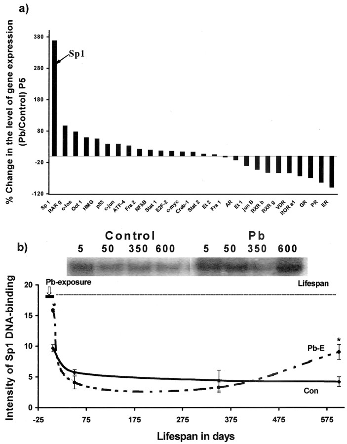
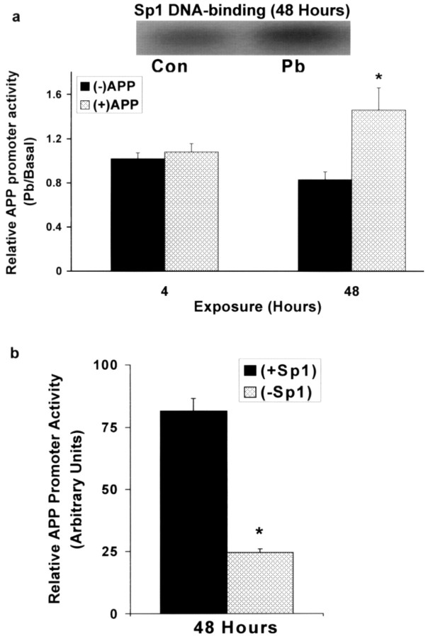
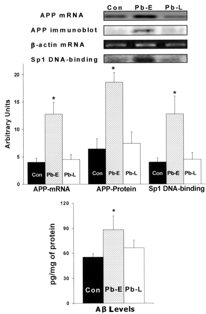
The fetal basis of adult disease (FeBAD) hypothesis states that many adult diseases have a fetal origin. According to FeBAD, injury or environmental influences occurring at critical periods of organ development could result in "programmatic" changes via alterations in gene expression or gene imprinting that may result in functional deficits that become apparent later in life. Alzheimer's disease (AD) is a progressive neurodegenerative disorder that is characterized by excessive deposits of aggregated beta-amyloid (Abeta) peptides, which are snippets of the beta-amyloid precursor protein (APP). The predominantly sporadic nature of AD suggests that the environment must play a role in neurodegeneration. To examine latent responses to an environmental agent, we exposed rodents to lead and monitored the lifetime expression of the APP gene. We observed that APP mRNA expression was transiently induced in neonates, but exhibited a delayed overexpression 20 months after exposure to Pb had ceased. This upregulation in APP mRNA expression was commensurate with a rise in activity of the transcription factor Sp1, one of the regulators of the APP gene. Furthermore, the increase in APP gene expression in old age was accompanied by an elevation in APP and its amyloidogenic Abeta product. In contrast, APP expression, Sp1 activity, as well as APP and Abeta protein levels were unresponsive to Pb exposure during old age. These data suggested that environmental influences occurring during brain development predetermined the expression and regulation of APP later in life, potentially altering the course of amyloidogenesis.


Figures

Figure 1.
Figure 1.
Lifetime changes in APP695 and β-actin mRNA expression in the cortex. Data shown are derived from two cohorts of animals separated in time by 1 year. The bar in the figure indicates the neonatal period of lactational exposure to Pb. Each data point represents the mean ± SEM for four to six animals. Data were analyzed by two-way ANOVA followed by a Duncan's post hoc test to compare the effects among various treatments; values marked with an asterisk are significantly different from their corresponding controls (p < 0.05).
Figure 2.
Figure 2.
mRNA profiling of transcription factors after developmental exposure to Pb and lifetime changes in Sp1 DNA-binding in the cortices of control and Pb-exposed animals. a, mRNA profiling. Cortical total RNA was isolated from P5 rats and used to probe cDNA arrays of transcription factors: AR, androgen receptor; ATF-4, activating transcription factor-4; c-fos, c-jun, c-myc, oncogenes; Creb-1, cAMP-responsive element-binding protein-1; E2F-2, ubiquitin-conjugating enzyme F-2; ER, estrogen receptor; Et, erythroblastosis virus oncogene homology; GR, glucocorticoid receptor; HMG, high-mobility group protein; NFκB, nuclearfactor κB; Oct 1, octamer transcription factor 1; p53, phosphoprotein 53; PR, progesterone receptor; RAR, retinoic acid receptor [related factors: retinoid orphan receptor (ROR), retinoid X receptor (RXR)]; Stat, signal transducer and activator of transcription; VDR, vitamin D3 receptor. The array data shown were derived from two sets of experiments, and each set consisted of tissue pooled from three animals. b, Sp1 DNA-binding. Nuclear extracts were prepared from controls and animals exposed as indicated by the bar in the figure. Sp1 DNA-binding was analyzed using gel mobility shift assays. Sp1 DNA-binding data shown are derived from two cohorts of animals separated in time by 1 year; each data point represents the mean ± SEM for four to six animals. Data were analyzed by two-way ANOVA followed by a Duncan's post hoc test to compare the effects among various treatments. Values marked with an asterisk are significantly different from their corresponding controls (p < 0.05).
Figure 3.
Figure 3.
Human APP promoter responsiveness after exposure to Pb and silencing of the Sp1 gene using siRNA in PC12 cells. a, Responsiveness of the APP promoter and Sp1 DNA-binding to Pb exposure. Con, Control. b, Silencing of the Sp1 gene. Cells were incubated in the presence or absence of Pb (0.1 μm) or siRNA (to knock out the Sp1 gene) and harvested to determine reporter gene activity or Sp1 DNA-binding. APP promoter activity plotted above is the ratio of activity of cells that contain pGL3 with the APP promoter versus cells transfected with pGL3 minus the APP promoter. Both cells contain the negative pRLTK vector. Promoter values shown are the means ± SEM of five to seven determinations. Data were analyzed by two-way ANOVA followed by a Duncan's post hoc test to compare the effects among various treatments; values marked with an asterisk are significantly different from their corresponding controls (p < 0.05).
Figure 4.
Figure 4.
Unresponsiveness of APP expression, Sp1 DNA-binding, APP and Aβ formation to Pb exposure in old age. Brain cortices were obtained from control 20-month-old animals and those directly exposed to 200 ppm of Pb-acetate as follows: control (Con, unexposed), Pb-E (exposed from P1 to P20), and Pb-L (exposed from 18 to 20 months of age). These tissues were used to analyze the mRNA expression of APP, β-actin (RT-PCR), Sp1 DNA-binding (gel shift), APP protein levels (Western blot), and Aβ activity (ELISA). Data shown are derived from two cohorts of animals separated in time by 1 year; each data point represents the mean ± SEM for four to six animals. Data were analyzed by two-way ANOVA followed by a Duncan's post hoc test to compare the effects among various treatments; values marked with an asterisk are significantly different from their corresponding controls (p < 0.05).

References

    1. Atwood CS, Moir RD, Huang X, Scarpa RC, Bacarra NM, Romano DM, Hartshorn MA, Tanzi RE, Bush AI (1998) Dramatic aggregation of Alzheimer abeta by Cu(II) is induced by conditions representing physiological acidosis. J Biol Chem 273: 12817-12826. - PubMed
    1. Bakheet SA, Zawia NH (2004) The role of pou domain transcription factors in lead neurotoxicity. In: Molecular neurotoxicology, environmental agents, and transcription-transduction coupling (Zawia NH, ed), pp 183-198. Boca Raton, FL: CRC.
    1. Barker DJ (2002) Fetal programming of coronary heart disease. Trends Endocrinol Metab 13: 364-368. - PubMed
    1. Barker DJ, Winter PD, Osmond C, Margetts B, Simmonds SJ (1989) Weight in infancy and death from ischaemic heart disease. Lancet 2: 577-580. - PubMed
    1. Barone Jr S, Stanton ME, Mundy WR (1995) Neurotoxic effects of neonatal triethyltin (TET) exposure are exacerbated with aging. Neurobiol Aging 16: 723-735. - PubMed

Publication types