. 2005 Jan 26;25(4):880-8.
doi: 10.1523/JNEUROSCI.4365-04.2005.

A novel substrate of receptor tyrosine phosphatase PTPRO is required for nerve growth factor-induced process outgrowth

Affiliations

A novel substrate of receptor tyrosine phosphatase PTPRO is required for nerve growth factor-induced process outgrowth

Bo Chen et al. J Neurosci. .

Abstract

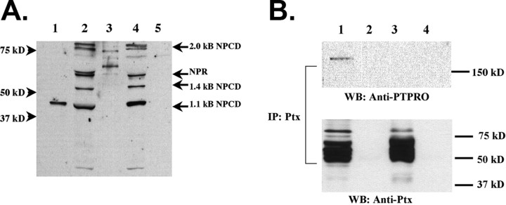
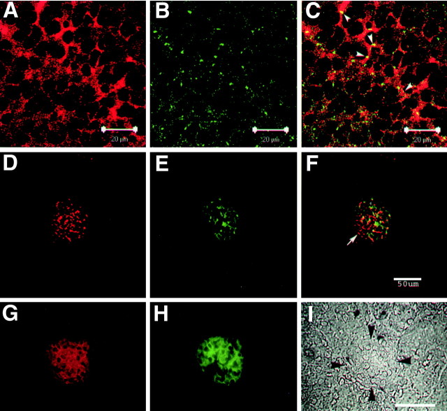
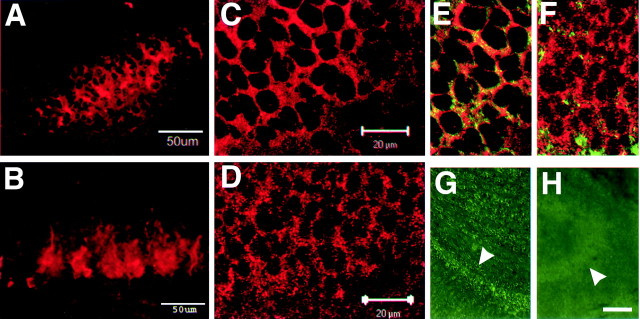
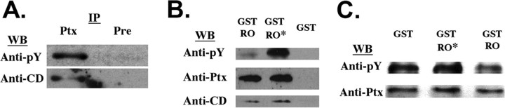
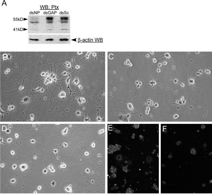
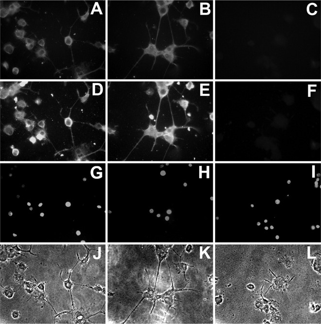
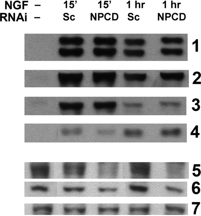
The receptor protein tyrosine phosphatase PTPRO may be involved in axon guidance both as a ligand and as a neuronal receptor. We have begun to characterize signaling by PTPRO as a receptor by screening for proteins interacting with the intracellular domain of PTPRO. In a yeast-two hybrid screen, we identified a novel class of protein, which we named neuronal pentraxin with chromo domain (NPCD), as a PTPRO-interacting protein. We have shown recently that NPCD has multiple cytoplasmic isoforms as a result of alternative splicing and that these proteins are present in many neurons, mainly associated with the inner side of the plasma membrane. Through additional two-hybrid experiments, cotransfection and reciprocal coprecipitation, glutathione S-transferase pulldown, and immunoprecipitation in vivo, we confirm that NPCD isoforms interact with the catalytic phosphatase domain of PTPRO. We also find that at least one NPCD isoform is tyrosine phosphorylated in vivo and can serve as a substrate for PTPRO in vitro. Analysis of PTPRO knock-out mice demonstrates that normal localization of NPCD at the plasma membrane requires PTPRO expression, suggesting a physiological role for the NPCD/PTPRO interaction. NPCD is likely to be relevant to axon growth and/or guidance, because RNA interference mediated knock-down of NPCD expression in pheochromocytoma cells inhibits NGF-induced neuronal process outgrowth without affecting NGF-dependent survival or initial NGF signaling.


Figures

Figure 1.
Figure 1.
PTPRO interacts with multiple NPCD isoforms and is present in a complex with NPCD in neonatal brain. A, The intracellular domain of PTPRO was produced as a GST fusion protein. Pulldowns were performed with GST-PTPRO (lanes 2, 4) or GST alone (lanes 3, 5) using either neonatal (lanes 2, 3) or adult (lanes 4, 5) mouse brain lysates. NPCD proteins were detected in the pulldowns by immunoblotting with an antibody recognizing a common epitope present in the NPCD pentraxin domain (a generous gift from Dr. Mark Perin). Multiple bands (41, 52, 55, 76 kDa) of sizes predicted from NPCD cDNAs (labeled arrows) were specifically precipitated by GST-PTPRO from either neonatal or adult brain. The 41 kDa NPCD protein produced in COS cells transfected with the 1.1 kb NPCD cDNA (lane 1) comigrates with one precipitated band. B, Immunoprecipitations were performed using our Ptx antibody (lanes 1, 3) or preimmune serum (lanes 2, 4) from the soluble fraction of either neonatal (lanes 1, 2) or adult (lanes 3, 4) brain lysates. Western blotting (WB) with anti-PTPRO revealed that PTPRO specifically coprecipitates from neonatal brain with NPCD. PTPRO could not be detected in the NPCD precipitates from adult brain (lane 3), although similar amounts of NPCD were precipitated. This result is likely attributable to downregulation of PTPRO expression in the adult brain. Note that the spectrum of NPCD species apparent in the anti-Ptx precipitate is a subset of those pulled down by the GST-PTPRO fusion protein.
Figure 2.
Figure 2.
NPCD colocalizes with PTPRO in brain and kidney. P0 mouse hippocampal sections (A-C) and adult kidney sections (D-F) were doubly immunostained with anti-Ptx (A,D) and anti-PTPRO (B,E), and confocal analysis (0.5 μm optical sections) was performed. Overlays are shown in C and F. Ptx-containing isoforms of NPCD appear localized mainly to the plasma membrane in CA3 hippocampal pyramidal neurons, where there is clear overlap (arrowheads, yellow; C) but not complete colocalization with PTPRO. Coexpression is present in the kidney, but there is extensive NPCD staining not associated with PTPRO (arrow, F). Conventional fluorescence microscopy of adult kidney sections (G-I) shows that both NPCD staining (red; G) and PTPRO staining (green; H) are confined to glomeruli (bordered by arrowheads in phase-contrast image; I) in adult kidney (Beltran etal., 2003). Scalebars: A-C,20 μm; F, I,50 μm.
Figure 3.
Figure 3.
PTPRO expression is required for appropriate localization of NPCD. A-F, Hippocampal sections from the CA3 region of P0 wild-type (A, C, E) and PTPRO null (B, D, F) mice were stained with anti-Ptx (A-D) or double stained with anti-Ptx and anti-NCAM (E, F) and examined by confocal fluorescence microscopy. A, B, Extended projections of 18 μm image stacks show that NPCD staining is less organized and appears less tightly associated with cell borders in PTPRO knock-out hippocampus (B) than in the wild type (A). Single optical sections show that NPCD is at or near the neuronal plasma membrane in wild-type hippocampus (C) but becomes more punctate and less tightly localized in the neurons of the PTPRO knock-out mice (D). The membrane association of NPCD in wild-type tissue can be assessed by general overlap with NCAM staining (green) in the same sections (E, F). Staining of wild-type (G) and PTPRO null hippocampus (H) with anti-PTPRO demonstrates the specificity of the punctate PTPRO staining in Figure 3. Arrowheads, Pyramidal layer. Scale bars: (in C, D) C-F,20 μm; H, 100 μm.
Figure 4.
Figure 4.
The 41 kDa NPCD protein is a PTPRO substrate. A, Immunoprecipitations were performed using the Ptx antibody (Ptx) or preimmune serum (Pre) from adult brain. The precipitates were probed with anti-phosphotyrosine (anti-pY) and were then stripped and reprobed with the CD antibody. An NPCD band at 41 kDa that is precipitated by the Ptx antibody and recognized by the CD antibody is tyrosine phosphorylated in vivo. B, GST pulldowns were performed from mouse brain lysates using GST-PTPRO (GST-RO), a catalytically inactive mutant of GST-PTPRO (GST-RO*), or GST alone. Precipitates were probed successively with a phosphotyrosine antibody, the Ptx antibody, and the CD antibody. Similar amounts of the 41 kDa NPCD protein were specifically associated with either the wild-type or inactive form of PTPRO. However, phosphotyrosine levels were much lower when the wild-type PTPRO was used, indicating that the 41 kDa NPCD isoform is a PTPRO substrate. C, Native 41 kDa NPCD protein was isolated from brain by immunoprecipitation using the Ptx antibody and was then incubated at 37°C for 1 h with 5 μg of purified GST, catalytically inactive GST-PTPRO, or GST-PTPRO. After incubation, NPCD was immunoprecipitated and probed successively with a phophotyrosine antibody and the Ptx antibody. NPCD is directly dephosphorylated by GST-PTPRO but not by the inactive mutant. WB, Western blot; IP, immunoprecipitation.
Figure 5.
Figure 5.
NPCD proteins are required for NGF-induced neuronal differentiation in PC12 cells. A, PC12 cells were transfected with siRNAs directed against NPCD (dsNP), GAPDH (dsGAP), or scrambled siRNAs (dsSc), and after 24 h, cell lysates were run on SDS-PAGE and sequentially blotted with anti-Ptx and anti-β-actin as a loading control. NPCD siRNAs severely reduced expression of the 55 kDa NPCD protein doublets (>90%) and caused a 40% reduction in expression of the 41 kDa NPCD protein compared with controls at this early time. B-F, PC12 cells were transfected with siRNAs that were scrambled so as not to match known sequences (B), siRNA capable of knocking down GAPDH (C, E), or siRNA for NPCD (D, F) and were then replated and grown in NGF for 3 d. Cells were examined by phase contrast (B-D) or fixed and stained for NPCD (E, F). Knock-down of NPCD was demonstrable after 4 d(F) and led to a lack of neurite growth (D). Hoechst staining of transfected cells revealed no apoptosis induced by NPCD siRNA (data not shown). These experiments were performed five times with similar results. Note that NPCD staining in PC12 cells requires permeabilization and is therefore cytosolic; the “ring” staining pattern suggests membrane association (Chen and Bixby, 2005). WB, Western blot.
Figure 6.
Figure 6.
NPCD proteins are required for neurite outgrowth of differentiated PC12 cells. Primed PC12 cells were treated with siRNAs for 3 d and were then replated onto poly-d-lysine-coated coverslips for an additional 12 h culture in the presence of NGF (100 ng/ml). Cells were treated with scrambled siRNAs (A, D, G, J), siRNAs directed against GAPDH (B, E, H, K), or siRNAs directed against NPCD (C, F, I, L). Staining of permeabilized cells with anti-Ptx (A-C) or anti-CD (D-F) demonstrates that NPCD protein expression is greatly reduced by NPCD siRNAs. Whereas primed PC12 cells treated with controls iRNA extended neurites within 12 h exposure to NGF (J, K), those treated with siRNAs directed against NPCD showed a minimum neurite growth response to NGF (L). Neurites were both shorter and less numerous after NPCD knock-down. There was no sign of apoptosis from siRNA treatment as shown by Hoescht-stained nuclei in the treated cells (G-I).
Figure 7.
Figure 7.
Initial NGF signaling through ERK and Akt pathways is unaffected by NPCD knock-down. PC12 cells were transfected with either scrambled (Sc) or NPCD siRNAs for 3 d before stimulation with NGF (100 ng/ml). Activation of signaling proteins was assessed 15 min and 1 h after NGF stimulation by Western blot with phospho-specific antibodies. We tested activation of ERKs 1 and 2 (row 1), the ERK target p90RSK (row 2), Akt (row 3), and the Akt target S6 ribosomal protein (row 4). No consistent effects on activation of any of these proteins at either time point was observed (a slight decrease seen here at 15 min for phosphoS6 was not seen in other experiments). NPCD knock-down of the 55 kDa doublet (row 5) and the 41 kDa band (row 6) were monitored in the same lysates; knock-down by NPCD siRNAs was clearly evident (85% knock-down compared with scrambled, 55 kDa; 60% knock-down, 41 kDa). GAPDH Western blotting was used in these blots as a loading control (row 7). This experiment was repeated twice with similar results. RNAi, RNA interference.

Similar articles

Cited by

References

    1. Ashcroft M, Stephens RM, Hallberg B, Downward J, Kaplan DR (1999) The selective and inducible activation of endogenous PI 3-kinase in PC12 cells results in efficient NGF-mediated survival but defective neurite out-growth. Oncogene 18: 4586-4597. - PubMed
    1. Beltran PJ, Bixby JL (2003) Receptor protein tyrosine phosphatases as mediators of cellular adhesion. Front Biosci 8: D87-D99. - PubMed
    1. Beltran PJ, Bixby JL, Masters BA (2003) Expression of PTPRO during mouse development suggests involvement in axonogenesis and differentiation of NT-3 and NGF-dependent neurons. J Comp Neurol 456: 384-395. - PubMed
    1. Bixby JL (2000) Receptor tyrosine phosphatases in axon growth and guidance. NeuroReport 11: R5-R10. - PubMed
    1. Bixby JL (2001) Ligands and signaling through receptor-type tyrosine phosphatases. IUBMB Life 51: 157-163. - PubMed

Publication types

MeSH terms