Comparative Study
. 2005 Jan 26;25(4):990-1001.
doi: 10.1523/JNEUROSCI.4388-04.2005.

Differential regulation of metabotropic glutamate receptor- and AMPA receptor-mediated dendritic Ca2+ signals by presynaptic and postsynaptic activity in hippocampal interneurons

Affiliations
Comparative Study

Differential regulation of metabotropic glutamate receptor- and AMPA receptor-mediated dendritic Ca2+ signals by presynaptic and postsynaptic activity in hippocampal interneurons

Lisa Topolnik et al. J Neurosci. .

Abstract

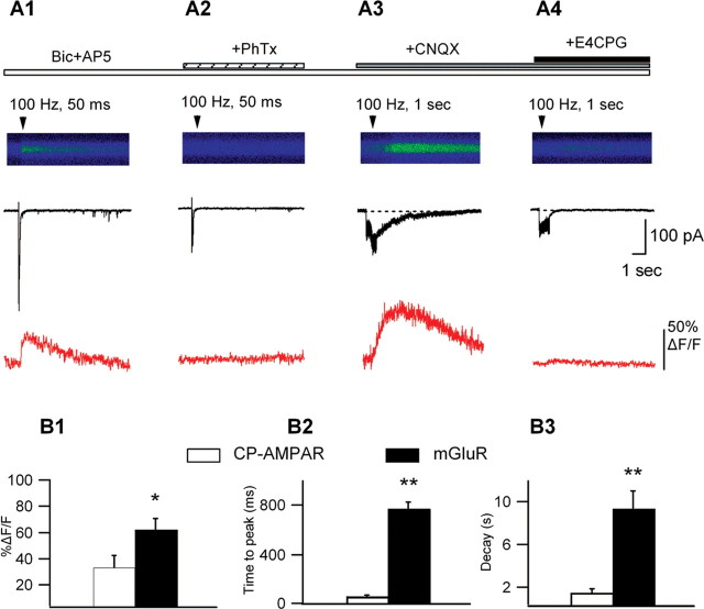
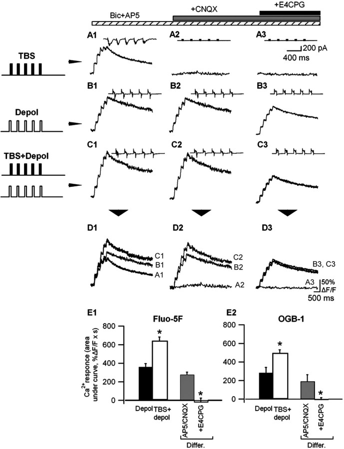
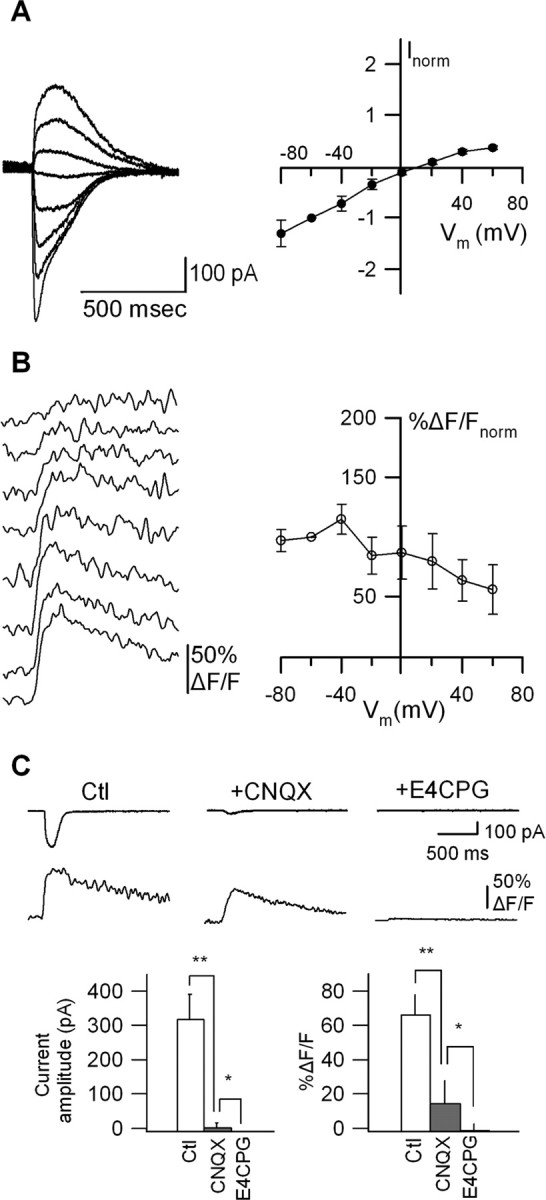
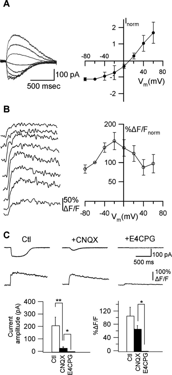
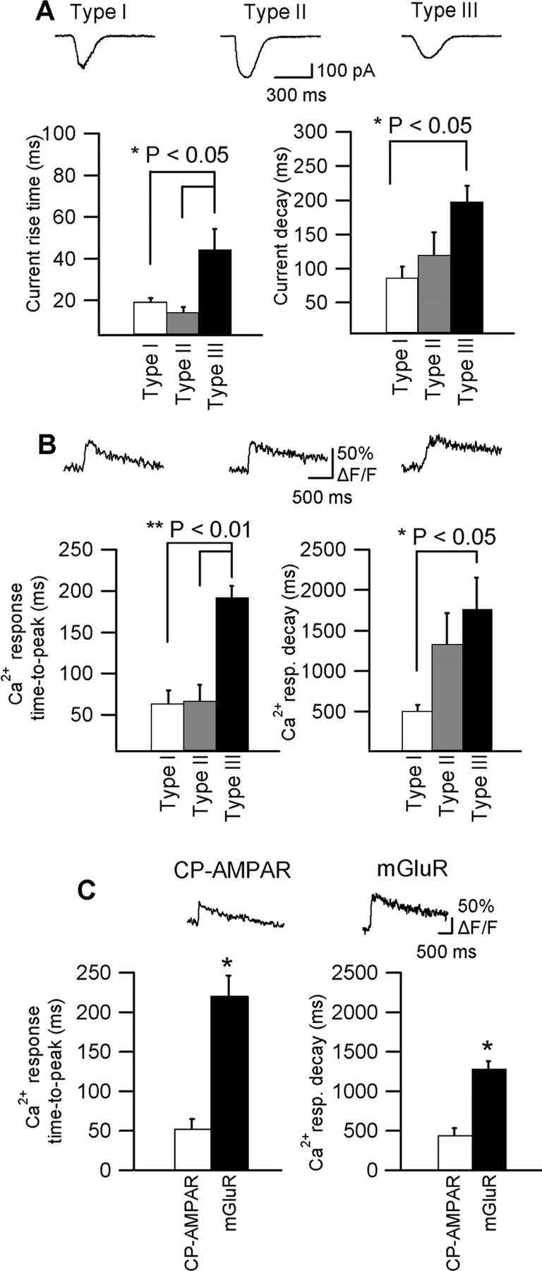
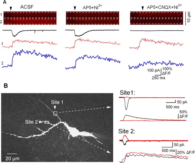
Calcium plays a crucial role as a ubiquitous second messenger and has a key influence in many forms of synaptic plasticity in neurons. The spatiotemporal properties of dendritic Ca2+ signals in hippocampal interneurons are relatively unexplored. Here we use two-photon calcium imaging and whole-cell recordings to study properties of dendritic Ca2+ signals mediated by different glutamate receptors and their regulation by synaptic activity in oriens/alveus (O/A) interneurons of rat hippocampus. We demonstrate that O/A interneurons express Ca2+-permeable AMPA receptors (CP-AMPARs) providing fast Ca2+ signals. O/A cells can also coexpress CP-AMPARs, Ca2+-impermeable AMPARs (CI-AMPARs), and group I/II metabotropic glutamate receptors (mGluRs) (including mGluR1a), in the same cell. CI-AMPARs are often associated with mGluRs, resulting in longer-lasting Ca2+ signals than CP-AMPAR-mediated responses. Finally, CP-AMPAR- and mGluR-mediated Ca2+ signals demonstrate distinct voltage dependence and are differentially regulated by presynaptic and postsynaptic activity: weak synaptic stimulation produces Ca2+ signals mediated by CP-AMPARs, whereas stronger stimulation, or weak stimulation coupled with postsynaptic depolarization, recruits Ca2+ signals mediated by mGluRs. Our results suggest that differential activation of specific glutamate receptor-mediated Ca2+ signals within spatially restricted dendritic microdomains may serve distinct signaling functions and endow oriens/alveus interneurons with multiple forms of Ca2+-mediated synaptic plasticity. Specific activation of mGluR-mediated Ca2+ signals by coincident presynaptic and postsynaptic activity fulfills the conditions for Hebbian pairing and likely underlies their important role in long-term potentiation induction at O/A interneuron synapses.


Figures

Figure 7.
Figure 7.
Differential regulation by presynaptic activity of synaptically elicited Ca2+ signals mediated by CP-AMPARs and mGluRs. A, Line scan images of synaptically evoked Ca2+ signals with representative currents (black) and associated Ca2+ transients (red) from the same cell in the presence of Bic and AP5 (A1), after PhTx block (A2), in the presence of Bic, AP5, and CNQX (A3), and after adding E4CPG (A4). Single bursts of high-frequency stimulation (30 μA; 5 stimuli at 100 Hz) produced summated EPSCs with appreciable Ca2+ influx (A1). These Ca2+ transients were completely blocked by PhTx (A2). In the absence of ionotropic glutamate transmission (in Bic, AP-5, and CNQX), repetitive synaptic stimulation (150 μA; 0.5-1 s at 100 Hz) evoked slower postsynaptic inward currents accompanied by slow Ca2+ transients (A3) that were both antagonized by E4CPG (A4). B, Bar graphs of the peak amplitude (B1), time-to-peak (B2), and decay time (B3) of synaptically evoked Ca2+ transients mediated by CP-AMPARs (PhTx sensitive; i.e., from A1) and by mGLuRs (E4CPG sensitive; i.e., from A3) (n = 5; *p < 0.05; **p < 0.01).
Figure 8.
Figure 8.
Regulation of Ca2+ signals mediated by mGluRs by postsynaptic depolarization. A1-C3, Representative Ca2+ transients measured with Fluo-5F and associated currents (insets) evoked by TBS at -60 mV (A1, A2), postsynaptic depolarization (-20 mV) (B1, B2), and TBS paired with depolarization (C1, C2) in different pharmacological conditions. D, Superimposed Ca2+ transients elicited in the same cell by the three stimulation protocols, showing that Ca2+ transients associated with TBS plus depolarization (C1, C2) were larger than those elicited by TBS (A1, A2) or depolarization (B1, B2) alone and that this difference was blocked by the mGluR antagonist E4CPG (B3, C3). E, Bar graphs of mean Ca2+ responses measured with Fluo-5F (E1) (n = 4 cells) and OGB-1 (E2) (n = 4 cells) evoked by depolarization alone (Depol), pairing TBS and depolarization (TBS + depol), and the difference in response between the two conditions (Differ.), showing that this difference was completely blocked by E4CPG (*p < 0.05).
Figure 1.
Figure 1.
Micropressure application of glutamate to interneuron dendrites produces local Ca2+ signals via multiple mechanisms. A, Multiphoton image (z-stack) of 200 optical sections taken at 0.5 μm intervals of an Oregon Green-filled interneuron; dendritic region of interest (ROI) near a puff-pipette is indicated by the box. B, Reconstruction of the same biocytin-filled interneuron. C, Pseudocolor images of an enlarged ROI with basal dendritic fluorescence (C1) and after glutamate puff (C2). C3, Dashed line indicates position for the line scan shown in D. D, Line scan images of the responses to glutamate puff (Glut. puff; C2). (arrowhead) with dashed lines bordering the region of interest for fluorescence measurements. Traces below are glutamate-evoked dendritic Ca2+ transients (red) and postsynaptic currents recorded from soma (black) (average of 4 sweeps). D2, Residual responses are the Ca2+ transient and membrane current independent of NMDARs and VGCCs [elicited in the presence of TTX (0.5 μm), Bic (10 μm), AP-5 (50 μm), and Ni2+ (50 μm)]. E, Spatiotemporal pattern of glutamate-evoked Ca2+ signals from a different cell; a dendritic ROI (E1), through which a line scan was placed, is indicated by the brackets. The amplitude of the calcium signal (ΔF/F) was measured from the corresponding line scan image (E2, top) and plotted as a function of distance along the dendrite for different times after glutamate application: 100 ms (red), 600 ms (green), 1100 ms (blue), and 1600 ms (black) (E2, bottom). These plots were well fitted by Gaussians. The x-axis of the plot (E2, bottom) corresponds to the length of the line scan image (E2, top). Note that in E2 (top) only the initial 1000 ms of the line scan is illustrated.
Figure 2.
Figure 2.
CP-AMPARs mediate dendritic Ca2+ transients associated with inwardly rectifying glutamate currents. A, Representative membrane currents (average of 4 sweeps) independent of NMDARs and VGCCs (in TTX, Bic, AP-5, Ni2+, and Cd2+) evoked by glutamate at different membrane potentials (Vm, -80 to +60 mV; left) and average I-V relationship for all cells (n = 5) with inward rectification (right). Before averaging, data from individual cells were normalized to the current amplitude at -60 mV. B, Associated Ca2+ transients (average of 4 sweeps) at different Vm (left) and plots of average peak Ca2+ transients for all cells as a function of Vm, showing steeply declining voltage dependence of Ca2+ transients (right). Data at different voltages were also normalized to Ca2+ transients at -60 mV. C, An example of use-dependent block of inwardly rectifying currents (left graph) by PhTx in one cell. Representative traces of membrane currents and associated Ca2+ transients before (Ctl) and after PhTx block are shown at right. D, Bar graph of the average amplitude of current and Ca2+ transient for all cells in the same conditions, showing that CP-AMPARs mediated this type of current and Ca2+ transients (n = 5; *p < 0.05).
Figure 3.
Figure 3.
Dendritic Ca2+ signals produced by joint activation of AMPARs/KARs and group I/II mGluRs. A, Representative glutamate-evoked membrane currents (average of 4 sweeps) at different Vm (left) and average I-V relationship for all cells (n = 6) showing intermediate inward rectification (right). B, Associated Ca2+ transients (average of 4 sweeps) at different Vm (left) and plots of average peak Ca2+ transients as a function of Vm, indicating slightly declining voltage dependence of Ca2+ transients (right). C, Representative currents and Ca2+ transients (top) evoked by glutamate puffs in control (Ctl) and in the presence of the AMPA/KAR antagonist (CNQX) and the group I/II mGluR antagonist (E4CPG), and bar graphs (bottom) of the average amplitude of current (left) and Ca2+ transient (right) in the same conditions. Currents and Ca2+ transients were sensitive to CNQX and E4CPG, indicating that a combination of AMPA/KARs and group I/II mGluRs participate in these responses (n = 6; *p < 0.05; **p < 0.01).
Figure 4.
Figure 4.
Dendritic Ca2+ signals mediated by group I/II mGluRs. A, Representative postsynaptic currents (average of 4 sweeps) at different Vm (left) and average I-V relationship for all cells (n = 14) with outward rectification (right). B, Associated Ca2+ transients (average of 4 sweeps) at different Vm (left) and plots of average peak Ca2+ transients for all cells as a function of Vm, showing bell-shaped voltage dependence of Ca2+ transients (right). C, Representative currents and Ca2+ transients evoked by glutamate puffs (top) in control (Ctl) and in the presence of the AMPA/KAR antagonist (CNQX) and the group I/II mGluR antagonist (E4CPG), and bar graphs (bottom) of the average amplitude of current (left) and Ca2+ transient (right) in the same conditions. Currents were sensitive to CNQX and E4CPG, whereas Ca2+ transients were significantly reduced only by E4CPG, showing that AMPA/KARs and group I/II mGluRs contribute to membrane currents, whereas only mGluRs mediate Ca2+ transients (n = 5; *p < 0.05; **p < 0.01).
Figure 5.
Figure 5.
Response-specific time course. A, Representative examples of three types of glutamate-evoked currents (Vh =-60 mV): type I (inwardly rectifying), type II (intermediate), and type III (outwardly rectifying), and bar graphs of the mean rise (left) and decay times (right) of currents for the three response types for all cells. B, Ca2+ transients associated with the three types of membrane currents shown in A and bar graphs of the mean time-to-peak (left) and decay time (right) of Ca2+ transients for all cells. Membrane currents and Ca2+ transients of type I and II responses displayed significantly faster rise time than those of type III response. Type III response showed significantly slower decay than type I response (**p < 0.01; *p < 0.05; ANOVA). C, Representative examples of CP-AMPAR-mediated (in AP-5 + E4CPG) and mGluR-mediated (in AP-5 + CNQX) Ca2+ transients measured with the low-affinity Ca2+ indicator Fluo-5F; bar graphs indicate the mean time-to-peak (left) and decay time (right) of Ca2+ transients for all cells (CP-AMPAR, n = 5; mGluR, n = 5).
Figure 6.
Figure 6.
Distinct Ca2+ signals in different dendritic sites of single interneurons. A, Representative line scan images of responses to glutamate applications (arrowhead). Dashed lines indicate the regions of interest for fluorescence measurements. Traces below illustrate glutamate-evoked currents at -60 mV (black) and associated dendritic Ca2+ transients measured within microdomains 1 (red) and 2 (blue). Distinct Ca2+ signals were produced in the two adjacent microdomains. At site 1 (red trace) the Ca2+ response was independent of NMDARs, VGCCs, and AMPA/KARs and was likely mediated by mGluRs, whereas at site 2 (blue trace) the Ca2+ response consisted of components mediated by NMDARs/VGCCs and AMPARs/KARs. B, Z-stack of an Oregon Green-filled interneuron (left) showing the two different locations of the puff-pipette and two dendritic regions of interest (1 and 2) indicated by the boxes. The corresponding membrane currents and Ca2+ transients evoked in each dendritic region at two Vm [Vh = -60 mV (black) and +40 mV (red)] are illustrated at right. The membrane currents at site 1 showed inward rectification, whereas those at site 2 showed an outward I-V relationship. The currents and Ca2+ transients are an average of four sweeps.

Similar articles

Cited by

References

    1. Baude A, Nusser Z, Roberts JD, Mulvihill E, McIlhinney RA, Somogyi P (1993) The metabotropic glutamate receptor (mGluR1 alpha) is concentrated at perisynaptic membrane of neuronal subpopulations as detected by immunogold reaction. Neuron 11: 771-787. - PubMed
    1. Bowie D, Mayer ML (1995) Inward rectification of both AMPA and kainate subtype glutamate receptors generated by polyamine-mediated ion channel block. Neuron 15: 453-462. - PubMed
    1. Carmant L, Woodhall G, Ouardouz M, Robitaille R, Lacaille JC (1997) Interneuron-specific Ca2+ responses linked to metabotropic and ionotropic glutamate receptors in rat hippocampal slices. Eur J Neurosci 9: 1625-1635. - PubMed
    1. Chuang SC, Bianchi R, Wong RK (2000) Group I mGluR activation turns on a voltage-gated inward current in hippocampal pyramidal cells. J Neurophysiol 83: 2844-2853. - PubMed
    1. Congar P, Ben-Ari Y, Crepel V (1997) A long-lasting calcium-activated nonselective cationic current is generated by synaptic stimulation or exogenous activation of group I metabotropic glutamate receptors in CA1 pyramidal neurons. J Neurosci 17: 5366-5379. - PMC - PubMed

Publication types

Substances

LinkOut - more resources