. 2005 Jan 26;25(4):1015-23.
doi: 10.1523/JNEUROSCI.3107-04.2005.

Polymodal sensory function of the Caenorhabditis elegans OCR-2 channel arises from distinct intrinsic determinants within the protein and is selectively conserved in mammalian TRPV proteins

Affiliations

Polymodal sensory function of the Caenorhabditis elegans OCR-2 channel arises from distinct intrinsic determinants within the protein and is selectively conserved in mammalian TRPV proteins

Irina Sokolchik et al. J Neurosci. .

Abstract

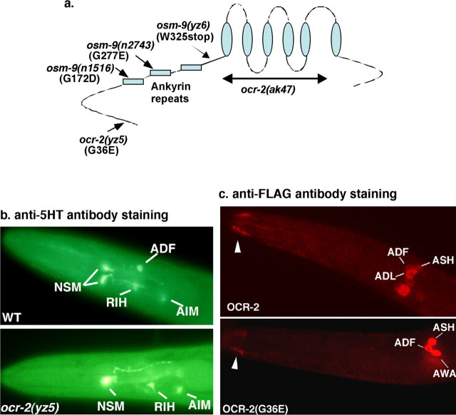
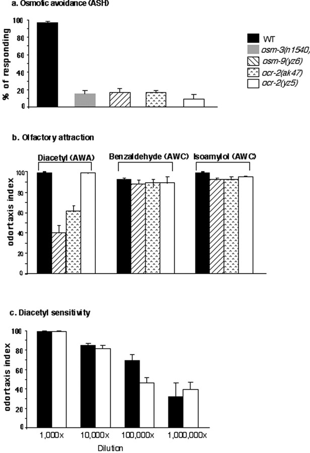
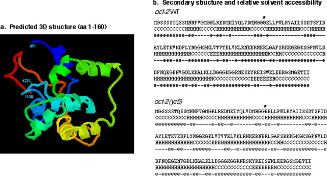
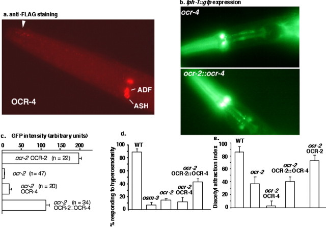
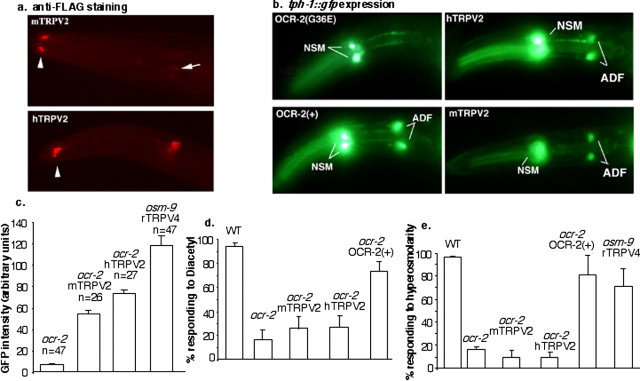
Caenorhabditis elegans OCR-2 (OSM-9 and capsaicin receptor-related) is a TRPV (vanilloid subfamily of transient receptor potential channel) protein that regulates serotonin (5-HT) biosynthesis in chemosensory neurons and also mediates olfactory and osmotic sensation. Here, we identify the molecular basis for the polymodal function of OCR-2 in its native cellular environment. We show that OCR-2 function in 5-HT production and osmotic sensing is governed by its N-terminal region upstream of the ankyrin repeats domain, but the diacetyl sensitivity is mediated by independent mechanisms. The ocr-2(yz5) mutation results in a glycine-to-glutamate substitution (G36E) within the N-terminal region. The G36E substitution causes dramatic downregulation of 5-HT synthesis in the ADF neurons, eliminates osmosensation mediated by the ASH neurons, but does not affect the response to the odorant diacetyl mediated by the AWA neurons. Conversely, wild-type sequence of the N-terminal segment confers osmotic sensitivity and upregulation of 5-HT production to a normally insensitive C. elegans homolog, OCR-4, but this chimeric channel does not respond to diacetyl stimuli. Furthermore, expression of either the mouse or human TRPV2 gene under the ocr-2 promoter can substantially restore 5-HT biosynthesis in ocr-2-null mutants but cannot improve the deficits in osmotic or olfactory sensation, suggesting that TRPV2 can substitute for the role of OCR-2 only in serotonergic neurons. Thus, different sensory functions of OCR-2 arise from separable intrinsic determinants, and specific functional properties of TRPV channel proteins may be selectively conserved across phyla.


Figures

Figure 1.
Figure 1.
The ocr-2(yz5) G36E substitution disrupts OCR-2 function in the serotonergic ADF neurons. a, Schematic representation of ocr-2 and osm-9 alleles used in this study. The ocr-2 and osm-9 proteins share the same domain structure and topology (Harteneck et al., 2000; Tobin et al., 2002). The ocr-2(ak47) and osm-9(yz6) mutations result in channel proteins lacking the transmembrane domain (indicated as cylinders) and are expected to be functional null alleles. It is noteworthy that the osm-9(n2743) and osm-9(n1516) mutations each result in a amino acid substitution in the ankyrin motifs and both cause deficits in every OSM-9/OCR-2 function (Colbert et al., 1997; Zhang et al., 2004), whereas the ocr-2(yz5) mutation discriminatively affects the functions. The position of amino acid changes corresponds, respectively, to the OSM-9 and OCR-2 protein sequences. b, Analysis of 5-HT immunoreactivity in WT and ocr-2(yz5) mutant animals. In WT, seven neurons of four classes in the head and two HSN neurons around the vulva (data not shown) are stained by the anti-5-HT antibody. In ocr-2(yz5) mutants, 5-HT immunoreactivity in the ADF neurons is dramatically reduced or absent, but 5-HT immunoreactivity in the other neurons is unaffected. Both ocr-2 and osm-9 are expressed in ADF but not in the other serotonergic neurons (Tobin et al., 2002; Zhang et al., 2004). Adult animals are shown, and the anterior is toward the left. c, Analysis of the expression pattern of FLAG-tagged OCR-2 and OCR-2(G36E). The FLAG epitope was tagged at the C terminus in both constructs. Consistent with the expression pattern revealed by ocr-2::gfp (Tobin et al., 2002), the immunofluorescence is predominantly observed in the cilia and plasma membrane of the ADF, ASH, ADL, and AWA neurons in the head and the PHA and PHB neurons in the tail; representative photomicrographs are shown. In general, the staining in the cilia is much weaker than in the plasma membrane, even with the OCR-2 construct. FLAG immunoreactivity of the OCR-2(G36E) fusion construct is lower overall, with a stronger effect observable in the cilia.
Figure 2.
Figure 2.
The ocr-2(yz5) G36E substitution selectively abrogates OCR-2 sensory functions. a, ocr-2(yz5) mutant animals fail to respond to osmotic stimuli. The assay tests for avoidance of 7 m glycerol. WT animals are repelled by high-osmolarity solutions and cannot escape from the ring of 7 m glycerol. osm-3 mutants have compromised sensory endings and presumably cannot sense; ∼85% of osm-3(n1540) mutants crossed the ring (Shakir et al., 1993). We repeatedly observed that higher percentages of ocr-2(yz5) mutants cross the ring than ocr-2(ak47), osm-9(yz6), or osm-3 mutants. b, ocr-2(yz5) mutants respond as well as WT to the odor attractant diacetyl. ocr-2(ak47) and osm-9(yz6) mutants exhibit profound deficits in sensing diacetyl at the dilution 1:1000. All of the mutants respond normally to benzaldehyde and isoamyl alcohol mediated by the AWC neurons. OCR-2 is not expressed in AWC, but it is expressed in AWA, which mediates diacetyl sensation. c, Comparison of diacetyl sensitivity between WT and ocr-2(yz5) mutant animals in series dilution assays. The neurons sensing the odorants are indicated. Each bar represents the mean ± SEM of at least three assays.
Figure 3.
Figure 3.
Structural model of the OCR-2 N-terminal segment. a, Prediction of the tertiary structure for OCR-2 (amino acids 1-160) (see Materials and Methods). G36 is predicted to reside within a fairly exposed coil between two α-helices. aa, Amino acids. b, Prediction of the secondary structure and relative solvent accessibility for wild-type OCR-2 and OCR-2(G36E) (amino acids 1-160) (see Materials and Methods). In wild-type OCR-2, G36 resides right before an α-helix. When G is changed to E, the E is predicted to become part of the helix. This shift could cause a global change in the side-chain phasing along the helix and alter intermolecular and intramolecular interactions. Notice the changes in the solvent accessibility of amino acids 33-60. The exposure of the residues was computed with a 30% threshold. The amino acid 36 is marked by asterisks. H, Helix; C, coil; e, exposed; —, buried.
Figure 4.
Figure 4.
The OCR-2 N-terminal region confers the regulation of tph-1 expression and osmotic sensation to normally insensitive OCR-4. a, Anti-FLAG antibody staining of ocr-2(ak47) mutants expressing a FLAG-tagged OCR-4 coding sequence under the promoter of ocr-2. The arrowhead points to the cilia of the chemosensory neurons. b, tph-1::gfp expression in ocr-2(ak47) mutants. The mutants bearing the OCR-2::OCR-4 chimeric channel recover tph-1::gfp expression, although the GFP level is not as high as in WT animals. OCR-4 on its own has no effect. c, Quantification of tph-1::gfp expression in the ADF neurons. All of the strains carry the same tph-1::gfp transgene. The fluorescence in a 25 × 25 pixel area in the ADF neurons was quantified by measuring the pixel intensity of the images captured using an Axiocam MR digital camera, as described in Materials and Methods. GFP intensity in multiple generations of the strains was examined. n, Number of animals examined. d, Osmotic response. ocr-2(ak47) mutant animals transformed with the OCR-2::OCR-4 chimeric channel partially restore the response to 7 m glycerol, whereas those transformed with OCR-4 are as defective as their nontransgenic siblings. Notice that OCR-2::OCR-4 exhibits an equivalent ability in rescuing osmotic sensation and tph-1::gfp expression of the ocr-2 mutants. Each bar represents the mean ± SEM of at least three assays. e, Diacetyl response. The OCR-2::OCR-4 chimeric channel does not improve diacetyl sensitivity of ocr-2(ak47) mutants, unlike the WT OCR-2 transgene. It is interesting to note that ocr-2(ak47) mutants bearing the OCR-4 transgene exhibited severer diacetyl sensory deficits than their nontransgenic sibling, perhaps attributable to an antimorph effect of OCR-4 in AWA function. Each bar represents the mean ± SEM of at least three assays.
Figure 5.
Figure 5.
Mouse and human TRPV2 genes recover tph-1::gfp expression in ocr-2(ak47) mutants. a, Anti-FLAG antibody staining. FLAG-tagged mouse and human TRPV2 are localized to the cilia and plasma membrane of OCR-2-expressing chemosensory neurons. The arrowheads point to the cilia. Although the mammalian TRPV2s and OCR-2 display a very similar expression pattern and subcellular location, the TRPV2 proteins tend to be concentrated in the cilia, whereas OCR-2 is more concentrated in the cytoplasma membrane (see Fig. 1c). b, tph-1::gfp expression in ocr-2 mutants transformed with various TRPV transgenes. The transgene in each strain is indicated. ocr-2 mutants bearing the OCR-2(G36E) transgene show absence of tph-1::gfp expression in the ADF neurons. Mouse and human TRPV2 substantially recover tph-1::gfp expression in ADF. The GFP levels in animals transformed with the mouse and human TRPV2 is not as high as those transformed with the WT OCR-2 transgene. c, Quantification of tph-1::gfp expression in the ADF neurons. All of the strains carry the same tph-1::gfp transgene. The fluorescence in a 25 × 25 pixel area in the ADF neurons was quantified by measuring the pixel intensity of the images captured using an Axiocam MR digital camera, as described in Materials and Methods. GFP intensity in multiple generations of the strains was examined. n, Number of animals examined. d, e, Mouse and human TRPV2 cannot improve osmotic or diacetyl deficits in ocr-2(ak47) mutants. In contrast, the OCR-2 construct can rescue both osmotic and diacetyl defects. All of the transgenes are expressed under the control of the same ocr-2 promoter, and each has the FLAG epitope tagged at the C terminus of the coding region. For behavioral assays, the data represent the response of transgenic animals identified based on their transgenic marker. Each bar represents the mean ± SEM of at least three assays.

Similar articles

Cited by

References

    1. Allessandri-Haber N, Yeh JJ, Boyd AE, Parada CA, Chen X, Reichling DB, Levine JD (2003) Hypotonicity induces TRPV4-mediated nociception in rat. Neuron 39: 497-511. - PubMed
    1. Baldi P, Pollastri G (2003) The principled design of large-scale recursive neural network architectures-DAG-RNNs and the protein structure prediction problem. J Machine Learning Res 4: 575-602.
    1. Baldi P, Chauvin Y, Hunkapiller T, McClure MA (1994) Hidden Markov models of biological primary sequence information. Proc Natl Acad Sci USA 91: 1059-1063. - PMC - PubMed
    1. Baldi P, Brunak S, Frasconi P, Soda G, Pollastri G (1999) Exploiting the past and the future in protein secondary structure prediction. Bioinformatics 15: 937-946. - PubMed
    1. Bargmann CI, Horvitz HR (1991a) Chemosensory neurons with overlapping functions direct chemotaxis to multiple chemicals in C. elegans Neuron 5: 729-742. - PubMed

Publication types

MeSH terms

LinkOut - more resources