. 2005 Jan 26;25(4):1024-33.
doi: 10.1523/JNEUROSCI.3951-04.2005.

Target-specific regulation of synaptic amplitudes in the neocortex

Affiliations

Target-specific regulation of synaptic amplitudes in the neocortex

Junryo Watanabe et al. J Neurosci. .

Abstract

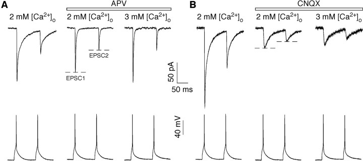
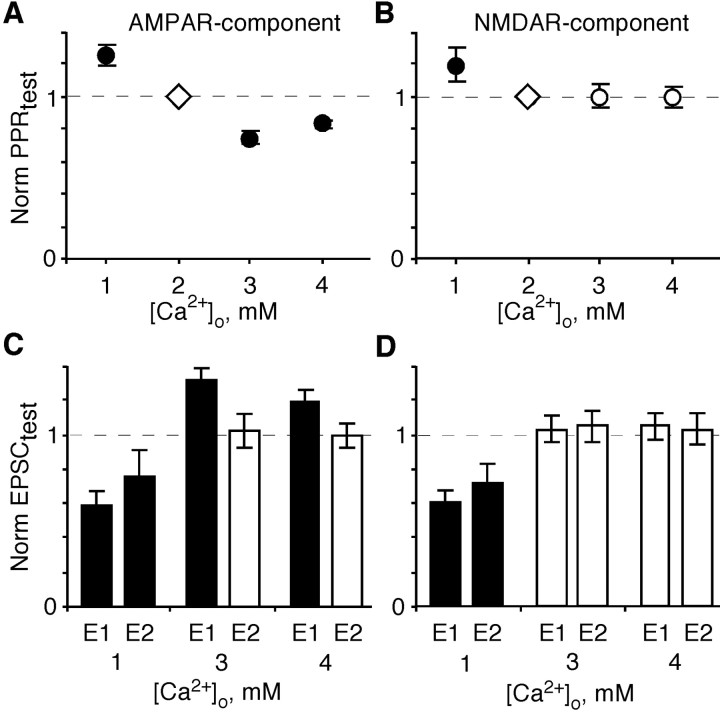
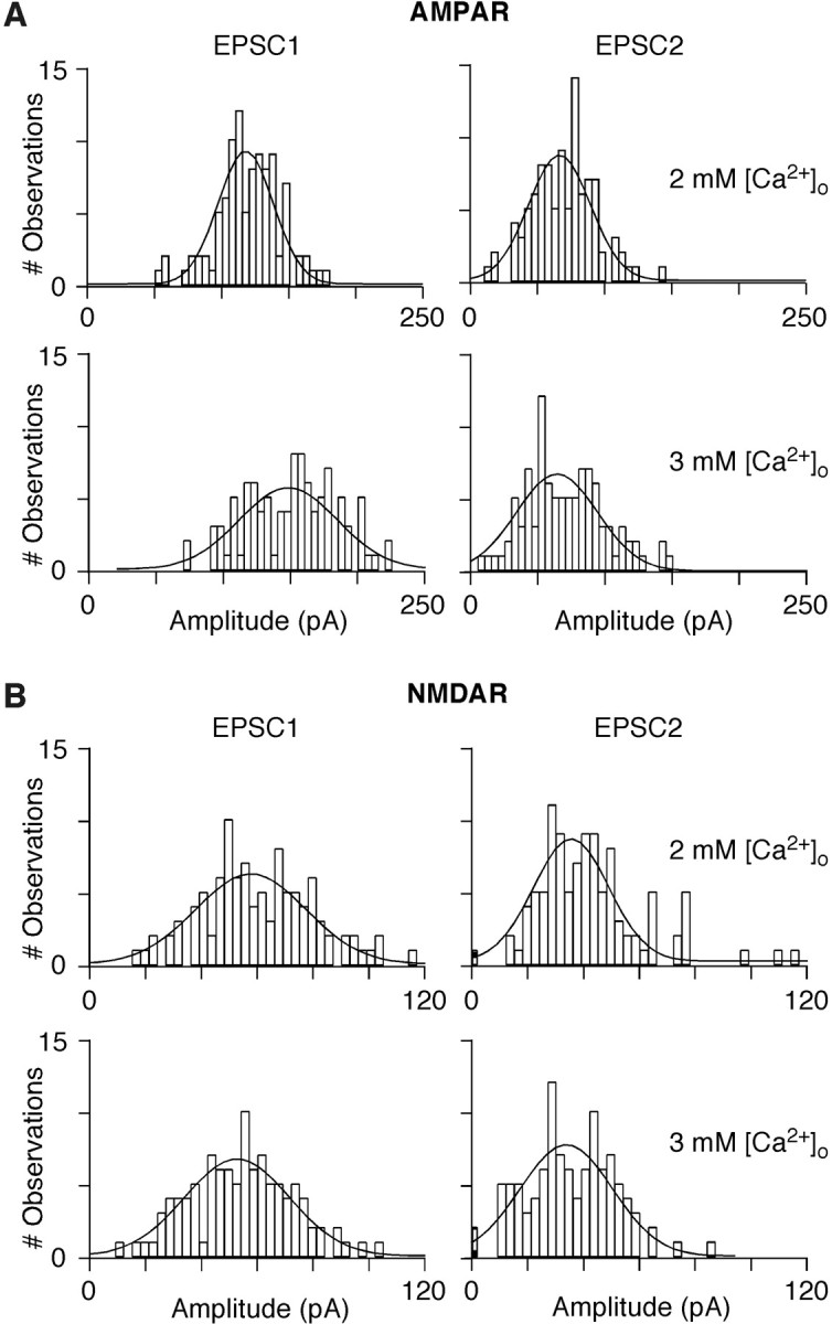
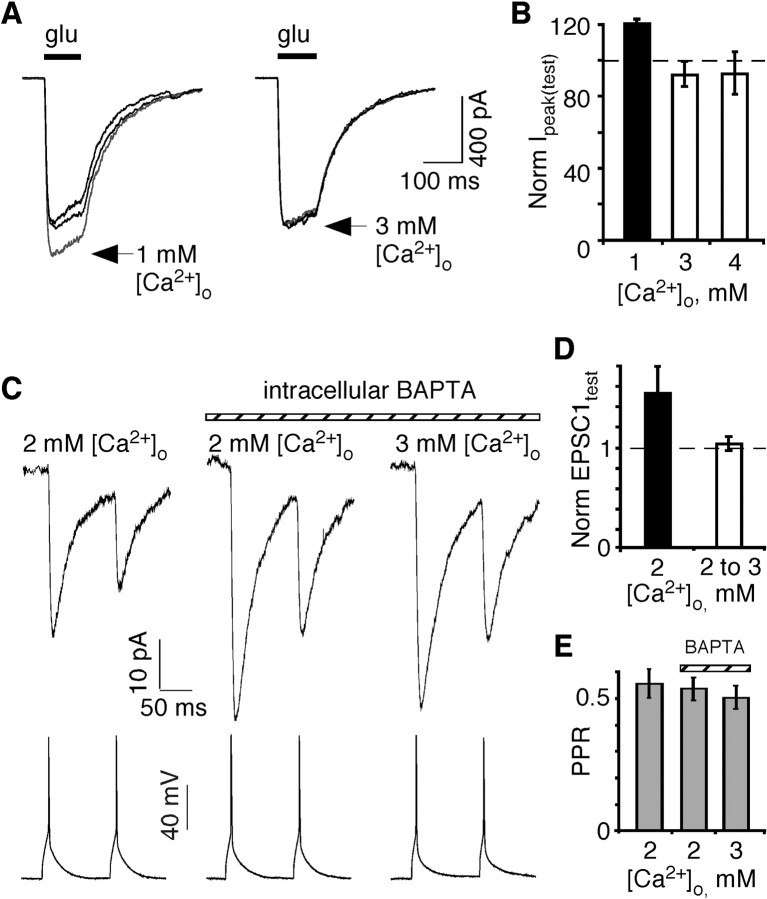
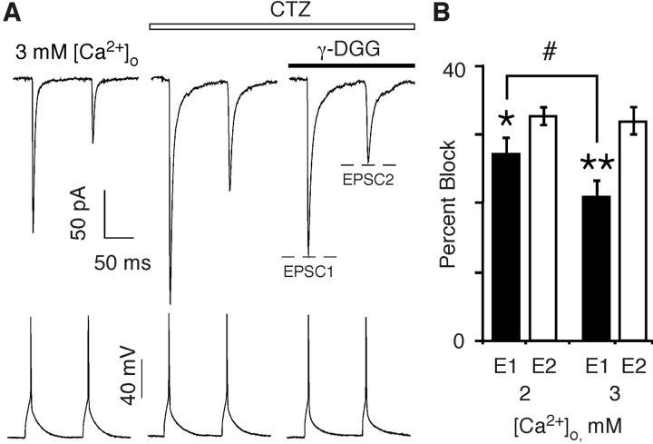
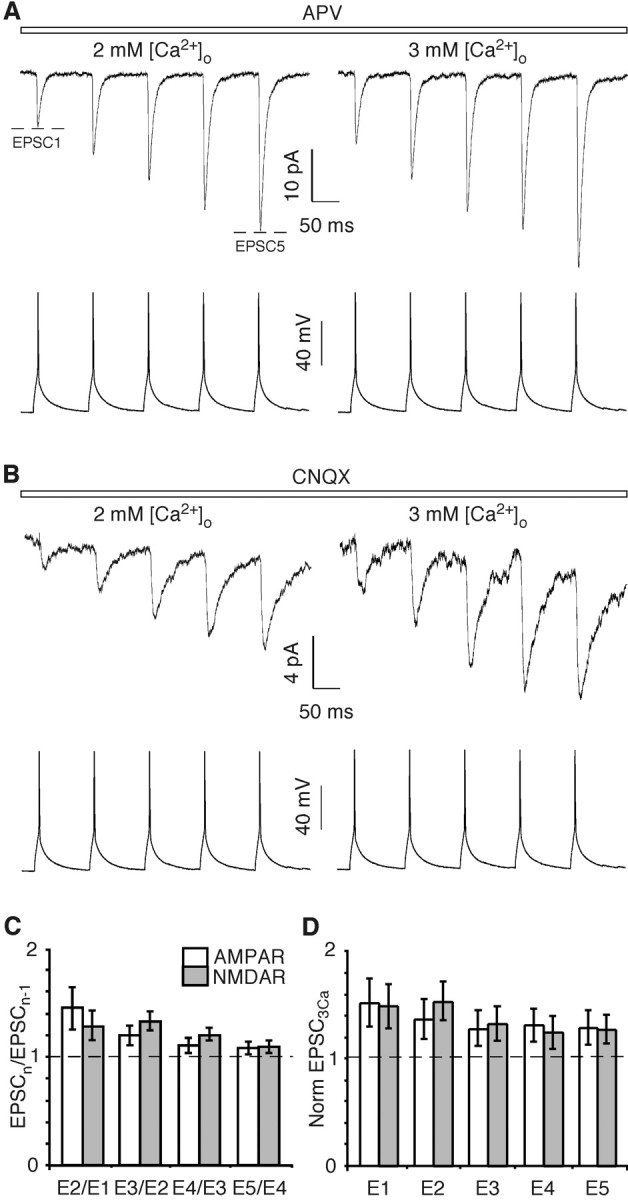
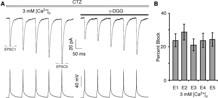
In layers 2/3 in the rat visual cortex, glutamatergic synapses, between pyramidal neurons and GABAergic interneurons, show target-specific depression or facilitation. To study the mechanisms regulating these short-term synaptic modifications, we recorded from synaptically connected pyramidal neurons (presynaptic) and multipolar or bitufted interneurons (postsynaptic). Evoked AMPA receptor (AMPAR)- or NMDA receptor (NMDAR)-mediated EPSCs were pharmacologically isolated at these pyramidal-to-interneuron synapses while altering release probability (P(r)) by changing the extracellular Ca2+ concentration ([Ca2+]o). At the pyramidal-to-multipolar synapse, which shows paired-pulse depression, elevation of [Ca2+]o from physiological concentrations (2 mm) to 3 mm increased the amplitude of the initial AMPAR-mediated EPSC and enhanced paired-pulse depression. In contrast, the initial NMDAR-mediated EPSC did not change in amplitude with raised P(r) nor was paired-pulse depression altered. This lack of an increase of NMDAR-mediated currents is not a result of Ca2+-dependent effects on the NMDAR. Rather, at the pyramidal-to-multipolar synapse, raised P(r) increases the transient glutamate concentration at individual release sites, possibly reflecting multivesicular release. In contrast, at the pyramidal-to-bitufted synapse, which shows facilitation, AMPAR- and NMDAR-meditated EPSCs showed parallel increases in response to raised P(r). Thus, our results reveal differential recruitment of AMPA and NMDARs at depressing and facilitating synapses in layers 2/3 of the cortex and suggest that the mechanisms regulating dynamic aspects of synaptic transmission are target specific.


Figures

Figure 1.
Figure 1.
AMPAR- and NMDAR-mediated EPSCs at the pyramidal-to-multipolar synapse. Simultaneous recordings of presynaptic pyramidal APs (bottom traces) and EPSCs from multipolar interneurons (top traces). EPSCs are an average of ∼100 sweeps. The far left trace in each panel was recorded in the absence of GluR antagonists. In the right two traces, the bathing solution contained either APV (A) (100 μm), an NMDAR competitive antagonist yielding the AMPAR-mediated component, or CNQX (B) (10 μm), an AMPAR competitive antagonist yielding the NMDAR-mediated component. These synapses do not have a significant kainate receptor component (Rozov et al., 2001a). The experimental paradigm was as follows: initial recordings were made in the 2 mm [Ca2+]o Ringer's solution with no GluR antagonists (left traces; control recording) (none of these solutions contained added MgCl2). Subsequently, recordings were made in the same solution with added APV or CNQX (middle traces; baseline recording) and then in 3 mm [Ca2+]o, APV, or CNQX (right traces; test recording) and finally back to 2 mm [Ca2+]o, APV, or CNQX (data not shown) (washout recording).
Figure 2.
Figure 2.
Changes in [Ca2+]o differentially altered PPR and EPSC amplitudes as monitored by AMPA and NMDARs. Comparison of the PPR (A, B) and the underlying EPSC amplitudes (C, D) (EPSC1, E1; EPSC2, E2) in a test [Ca2+]o (1, 3, or 4 mm) normalized to that recorded in the same pair in 2 mm [Ca2+]o. All experiments were performed as outlined in Figure 1, isolating either the AMPAR-mediated (A, C) or NMDAR-mediated (B, D) components. Number of pairs, from left to right, 6, 18, 7, 5 (A), and 7, 27, 10, 10 (B). In C and D, recordings are from the same set of pairs as shown in A and B. Solid symbols or bars in this and all subsequent figures indicate values significantly different from control or baseline responses.
Figure 3.
Figure 3.
Amplitude histograms of individual EPSCs mediated by AMPA and NMDARs. A comparison of individual EPSC amplitudes for pyramidal-to-multipolar pairs recorded in APV (A) or CNQX (B). Within a panel, the amplitudes for EPSC1 (left column) and EPSC2 (right column) are shown in 2 mm [Ca2+]o (top row) or in 3 mm [Ca2+]o (bottom row). Bin width is 5 pA for the AMPAR and 3 pA for the NMDAR component. Failures are shown in shaded boxes. Number of failures (amplitudes were within noise)/trials: 2 mm [Ca2+]o: EPSC1, 0/100; EPSC2, 0/100; 3 mm [Ca2+]o: EPSC1, 0/100; EPSC2, 0/100 (A); 2 mm [Ca2+]o, EPSC1, 0/104; EPSC2, 1/104; 3 mm [Ca2+]o: EPSC1, 0/104; EPSC2, 2/104 (B). Continuous lines represent Gaussian fits.
Figure 4.
Figure 4.
Effect of extracellular and intracellular Ca2+ on NMDAR in multipolar interneurons. A, Recordings of NMDAR-mediated currents in nucleated patches taken from presumed multipolar interneurons. Patches were bathed either in 2 mm [Ca2+]o (thick traces) before and after a recording in 1 mm (left) or 3 mm (right) (lighter traces) [Ca2+]o. Currents were elicited by glutamate (200 μm; horizontal bar) at a holding potential of -60 mV. In all instances, the external solution contained added glycine (10 μm) and CNQX (10 μm). B, Average relative peak current amplitudes in a test [Ca2+]o (1, 3, or 4 mm) (Ipeak(test)) normalized to that in 2 mm [Ca2+]o. Number of recordings, from left to right: 5, 5, 4. C, Simultaneous recordings of presynaptic pyramidal APs (bottom traces) and average EPSCs from multipolar interneurons (top traces) in the presence of CNQX (10 μm). The far left trace was recorded in the 2 mm [Ca2+]o Ringer's solution (without added MgCl2) using our standard (no Ca2+ buffer) pipette solution. Subsequently, the patch-pipette recording of the postsynaptic multipolar interneuron was replaced with one containing the same internal solution plus 2 mm BAPTA. After an ∼5 min wait period, EPSCs were initially recorded in 2 mm (center trace) and then in 3 mm (right trace) [Ca2+]o as in Figure 1 A. D, Amplitude of EPSC1 normalized to that in 2 mm [Ca2+]o. Left bar, EPSC1test was recorded in 2 mm [Ca2+]o with 2 mm BAPTA in the pipette and was normalized to records with 0 BAPTA in the pipette (n = 6). Right bar, EPSC1test was recorded in 3 mm [Ca2+]o and 2 mm BAPTA and normalized to records with 2 mm BAPTA in the pipette (n = 6). E, Paired-pulse ratio under the different recording conditions shown in C.
Figure 5.
Figure 5.
Differential effect of γ-DGG on AMPAR-mediated EPSCs. A, Average AMPAR-mediated EPSCs from multipolar interneurons (top traces) recorded in the 3 mm [Ca2+]o Ringer's solution (with added 1 mm MgCl2) either in the absence (left trace) or presence (right two traces) of CTZ (50 μm). γ-DGG (0.5 mm; far right trace) was applied in the presence of CTZ. B, Percentage block of EPSC1 and EPSC2 by γ-DGG recorded either in 2 or 3 mm [Ca2+]o and CTZ. Number of pairs, 2 mm [Ca2+]o (n = 9) and 3 mm [Ca2+]o (n = 8). The various symbols indicate significant differences (t test) in the percentage block between EPSC1 and EPSC2 in 2 mm [Ca2+]o (*p < 0.02), EPSC1 and EPSC2 in 3 mm [Ca2+]o (**p < 0.0009), or EPSC1 in 2 mm [Ca2+]o and EPSC1 in 3 mm [Ca2+]o (#p < 0.04).
Figure 6.
Figure 6.
Effect of increases in [Ca2+]o on AMPAR- and NMDAR-mediated EPSCs at the pyramidal-to-bitufted synapse. A, B, Simultaneous recordings of presynaptic pyramidal APs (bottom traces) and average AMPAR-mediated (A) and NMDAR-mediated (B) EPSCs from bitufted interneurons (top traces) in either 2 mm (left) or 3 mm (right) [Ca2+]o. Experimental protocol, solutions, and analysis are as in Figure 1, except that five presynaptic APs were stimulated (at 10 Hz). C, Ratio of the amplitude of successive AMPAR-mediated (n = 10) or NMDAR-mediated (n = 8) EPSCs. Recordings were made in 2 mm [Ca2+]o. All ratios were significantly greater than unity, with the AMPAR-mediated E2/E1 ratio significantly different from the others. D, AMPAR- and NMDAR-mediated EPSC amplitudes in 3 mm [Ca2+]o normalized to that recorded in the same pair in 2 mm [Ca2+]o. All ratios were significantly greater than unity but were not significantly different from each other.
Figure 7.
Figure 7.
Effect of γ-DGG on AMPAR-mediated EPSC amplitudes. A, Average EPSCs from bitufted interneurons (top traces) recorded in the 3 mm [Ca2+]o Ringer's solution (with added 1 mm MgCl2). All shown traces were recorded in the presence of CTZ (50 μm). γ-DGG (0.5 mm; right trace) was applied in the presence of CTZ. B, Percentage block of EPSCs by γ-DGG recorded in 3 mm [Ca2+]o and CTZ (n = 7). None of the values were significantly different.

Similar articles

Cited by

References

    1. Abbott LF, Varela JA, Sen K, Nelson SB (1997) Synaptic depression and cortical gain control. Science 275: 220-224. - PubMed
    1. Alger BE (2002) Retrograde signaling in the regulation of synaptic transmission: focus on endocannabinoids. Prog Neurobiol 68: 247-286. - PubMed
    1. Arrigoni E, Rainnie DG, McCarley RW, Greene RW (2001) Adenosine-mediated presynaptic modulation of glutamatergic transmission in the laterodorsal tegmentum. J Neurosci 21: 1076-1085. - PMC - PubMed
    1. Auger C, Kondo S, Marty A (1998) Multivesicular release at single functional synaptic sites in cerebellar stellate and basket cells. J Neurosci 18: 4532-4547. - PMC - PubMed
    1. Cauli B, Audinat E, Lambolez B, Angulo MC, Ropert N, Tsuzuki K, Hestrin S, Rossier J (1997) Molecular and physiological diversity of cortical nonpyramidal cells. J Neurosci 17: 3894-3906. - PMC - PubMed

Publication types

MeSH terms