. 2005 Feb 24;24(9):1491-500.
doi: 10.1038/sj.onc.1208307.

Induction of KLF4 in basal keratinocytes blocks the proliferation-differentiation switch and initiates squamous epithelial dysplasia

Affiliations

Induction of KLF4 in basal keratinocytes blocks the proliferation-differentiation switch and initiates squamous epithelial dysplasia

K Wade Foster et al. Oncogene. .

Abstract

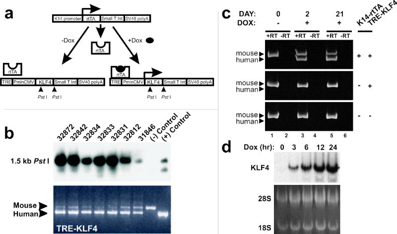
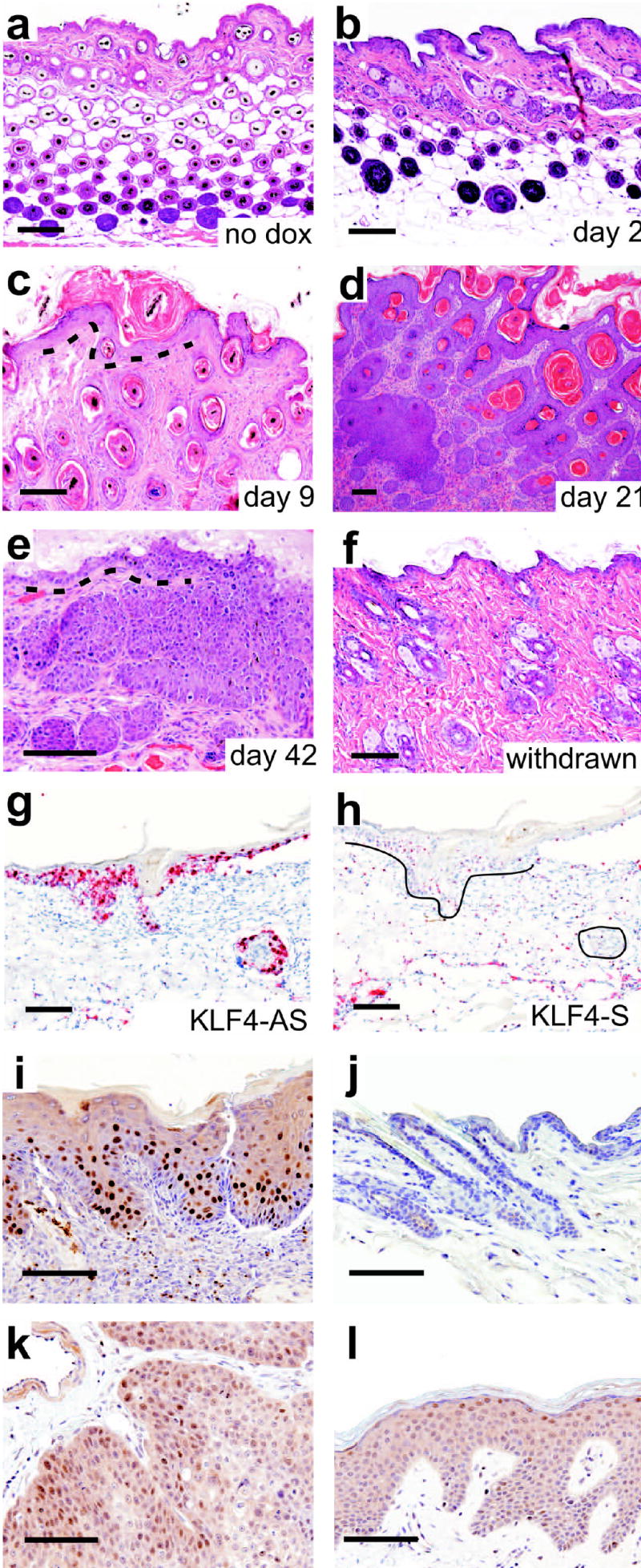
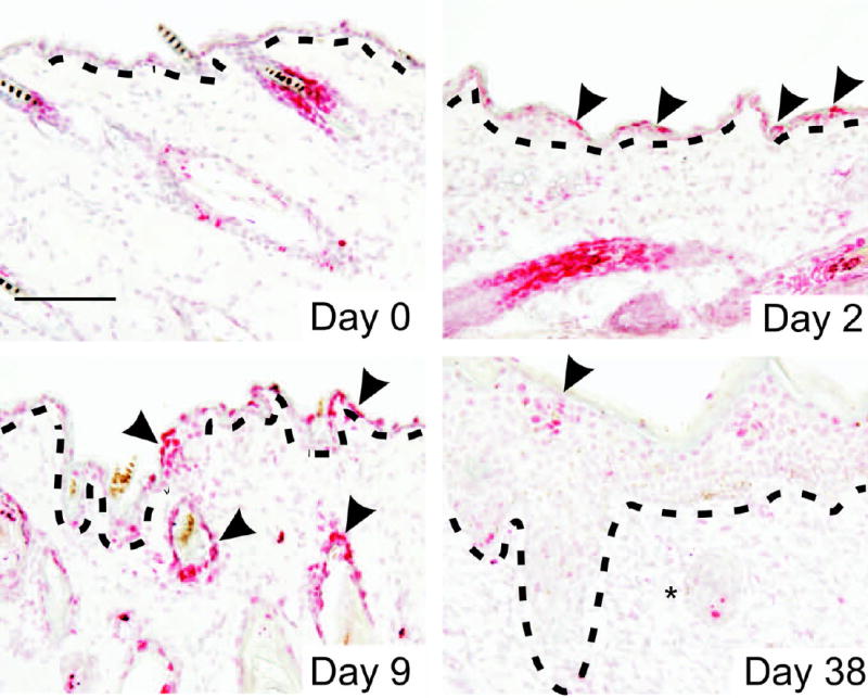
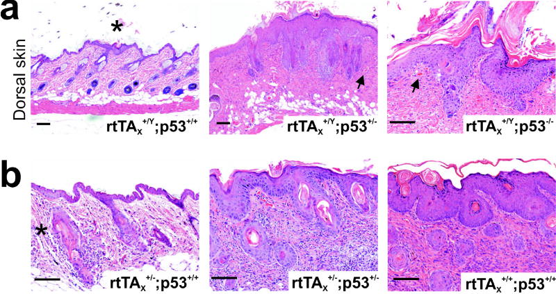
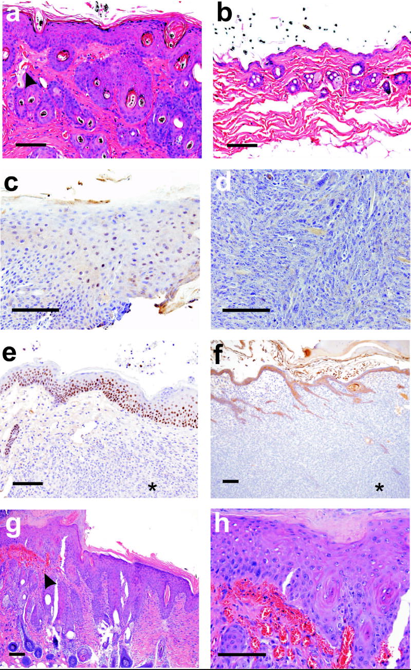
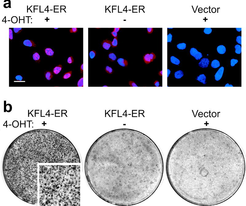
KLF4/GKLF normally functions in differentiating epithelial cells, but also acts as a transforming oncogene in vitro. To examine the role of this zinc finger protein in skin, we expressed the wild-type human allele from inducible and constitutive promoters. When induced in basal keratinocytes, KLF4 rapidly abolished the distinctive properties of basal and parabasal epithelial cells. KLF4 caused a transitory apoptotic response and the skin progressed through phases of hyperplasia and dysplasia. By 6 weeks, lesions exhibited nuclear KLF4 and other morphologic and molecular similarities to squamous cell carcinoma in situ. p53 determined the patch size sufficient to establish lesions, as induction in a mosaic pattern produced skin lesions only when p53 was deficient. Compared with p53 wild-type animals, p53 hemizygous animals had early onset of lesions and a pronounced fibrovascular response that included outgrowth of subcutaneous sarcoma. A KLF4-estrogen receptor fusion protein showed tamoxifen-dependent nuclear localization and conditional transformation in vitro. The results suggest that KLF4 can function in the nucleus to induce squamous epithelial dysplasia, and indicate roles for p53 and epithelial-mesenchymal signaling in these early neoplastic lesions.


Figures

Figure 1
Figure 1
Doxycycline (dox) inducible KLF4 transgenic mouse lines. (a) A transgene composed of the keratin 14 (K14) promoter and the reverse tetracycline-responsive transcriptional activator (rtTA) directs expression of tetracycline response element (TRE)-linked transgenes to K14 positive cell types. Other elements of the TRE-KLF4 transgene include the minimal cytomegalovirus promoter (PminCMV) and an intron (Int) and polyadenylation signal (poly A) from the SV40 genome. (b) Upper panel: Southern analysis of transgenic mouse lines was performed using a human KLF4 cDNA probe derived from the 3′ UTR. Lower panel: PCR analysis used conserved primers derived from different exons to co-amplify mouse genomic DNA and the human transgene. Control DNAs were mouse genomic DNA alone or else mouse DNA admixed with a molar excess of KLF4 cDNA. (c) RT-PCR analysis of KLF4 in mouse skin. The mouse and human PCR products differ in size by 20bp. Genotypes are indicated on the right. (d) Primary keratinocytes were prepared from K14-rtTA;TRE-KLF4 newborn mice (Dlugosz et al., 1995). Dox was added to the culture media for the indicated interval, and total RNA was analyzed by Northern. Ethidium bromide-stained RNA was visualized by irradiation of the filter (below).
Figure 2
Figure 2
Histology of the skin following induction of KLF4. (a–f) Dox was administered for the indicated interval and ventral skin of male animals was analyzed. The dermo-epidermal junction (DEJ) is marked with a dashed line (c, e). In parallel with these studies, dox was withdrawn between days 21 and 42 (f). (g–h) mRNA in situ hybridization analysis of dysplastic skin lesions. Antisense (AS) or sense control (S) probes for KLF4 were applied to serial sections. Epithelial-mesenchymal boundaries are indicated (panel h, black lines). Scale bar, 100μ. (i–l) Analysis of KLF4 using affinity-purified, polyclonal antibody (anti-KLF4). (i) Dysplastic mouse skin after 4 weeks of induction. (j) Nontransgenic skin was a control. (k–l) Adjacent areas of a human tissue section contained cutaneous SCC (k) or mild dysplasia (l). Scale bar, 100μ.
Figure 3
Figure 3
Immunostaining of KLF4-induced lesions. The indicated antibodies were applied to sections of ventral skin of males. Arrowheads indicate the DEJ. Asterisks indicate lesions deeper within the dermis that are PCNA-positive and K1-low, similar to human SCC. Scale bar, 100μ.
Figure 4
Figure 4
TUNEL analysis of apoptosis following induction of KLF4. Frozen sections were labeled with digoxigenin (dig)–dUTP and terminal transferase. Dig was detected using an alkaline phosphatase conjugated antibody and the substrate Fast Red (arrowheads). At 38 days, dysplastic epithelium exhibited TUNEL staining that was limited to the most superficial cells. Epithelial cells deeper within the dermis are indicated with an asterisk. Sections were lightly counterstained with hematoxylin. The DEJ is indicated with a dashed line. Scale bar, 100μ.
Figure 5
Figure 5
p53 gene dosage alters the skin phenotype of TRE-KLF4 transgenic mice. (a) Analysis of dorsal skin in p53 wild-type (p53+/+) or deficient (p53+/−, p53−/−) males. p53+/+ animals exhibited only focal, minor involvement (left panel, asterisk). In contrast, p53+/− animals exhibited epithelial dysplasia and a prominent fibrovascular response that effaced several mm of the dermis (middle panel, 3 week induction). In the right panel, a p53−/− animal showed increased nuclear atypia following 4 weeks of induction. Blood vessels near the DEJ are indicated by arrows (middle and right panels). (b) Mosaic expression in female skin resulting from an X chromosome-linked rtTA transgene (rtTAX+/−) induced only focal, minor abnormalities (left panel, asterisk). Either p53 hemizygosity (middle panel) or uniform expression (rtTAX+/+, right panel) resulted in the dysplastic phenotype. The results shown were observed in each of 5 or more animals. Scale bars, 100μ.
Figure 6
Figure 6
Analysis of MMTV-KLF4 transgenic mice. (a) p53+/− animals developed dorsal hair loss, dysplasia, and fibrotic skin between 6 and 8 mo. of age in association with subcutaneous sarcoma. Dysplasia appeared similar to the inducible model, with a prominent fibrovascular response, and blood vessels near the DEJ (black arrowhead). (b) Skin of an MMTV-KLF4;p53+/− animal at 3 mo. of age. (c–f) Immunostaining of dysplasia and/or the associated sarcoma in an MMTV-KLF4;p53+/− animal. (c) Staining of dysplastic epithelium with anti-KLF4. (d) Absence of staining of adjacent sarcoma cells with anti-KLF4. (e) Increased nuclear staining by anti-p63 in epithelial cells vs. sarcoma cells (asterisk). (f) Differential staining of epithelium with anti-keratin AE1/AE3, and extensive intercalation of keratin-positive epithelium with the underlying sarcoma (asterisk). (g–h) Skin of an MMTV-KLF4;p53+/+ animal at 18 mo. of age. (g) Dysplasia with prominent vascularization and a fibrotic response near the DEJ (black arrowhead). (h) The region marked in panel g is shown at higher magnification, demonstrating nuclear pleomorphism, hyperchromicity, frequent mitoses, and blood vessels adjacent to the DEJ. Scale bars, 100μ.
Figure 7
Figure 7
Characterization of KLF4-ER. (a) KLF4 was fused to a 4-hydroxytamoxifen (4-OHT)-responsive fragment of the mouse estrogen receptor (ER). Stably-transduced cells were stained to enable localization of KLF4-ER. Nuclear localization results in a pink color in the merged red (antibody) and blue (nuclei) images. (b) KLF4-ER or Vector control retrovirus was transduced into a small subset of RK3E epithelial cells, and cultures were maintained in the presence of 4-OHT or vehicle. Dishes were stained to identify transformed foci (inset).

Similar articles

Cited by

References

    1. Adam PJ, Regan CP, Hautmann MB, Owens GK. J Biol Chem. 2000;275:37798–37806. - PubMed
    1. Arbeit JM, Munger K, Howley PM, Hanahan D. Journal of Virology. 1994;68:4358–4368. - PMC - PubMed
    1. Boyle JO, Hakim J, Koch W, van der RP, Hruban RH, Roa RA, Correo R, Eby YJ, Ruppert JM, Sidransky D. Cancer Res. 1993;53:4477–4480. - PubMed
    1. Brash DE, Ponten J. Cancer Surv. 1998;32:69–113. - PubMed
    1. Brown K, Balmain A. Cancer Metastasis Rev. 1995;14:113–124. - PubMed

Publication types

MeSH terms