

Review
doi: 10.1086/428361.
Epub 2005 Jan 24.
The D4Z4 repeat-mediated pathogenesis of facioscapulohumeral muscular dystrophy
Affiliations
- PMID: 15674778
- PMCID: PMC1196390
- DOI: 10.1086/428361
Item in Clipboard
Review
The D4Z4 repeat-mediated pathogenesis of facioscapulohumeral muscular dystrophy
Am J Hum Genet.
2005 Mar.
No abstract available
Figures
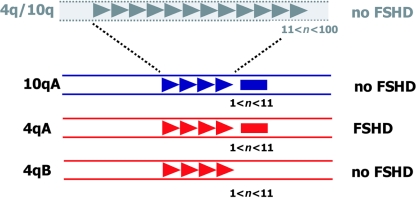
Schematic presentation of the primary genetic defect of FSHD. Polymorphic arrays of D4Z4 units (triangles) are located in the subtelomeres of chromosomes 4q (red) and 10q (blue). On chromosome 10, this array may vary between 1 and 100 units without pathological consequences (n = number of units). On chromosome 4, however, repeat arrays in the range of 1–10 units cause FSHD, but only when the contraction is in cis with the subtelomeric variant 4qA, which contains beta satellite repeats (red rectangle), not the 4qB variant. The beta satellite repeats are also present in the 10q subtelomere (blue rectangle).
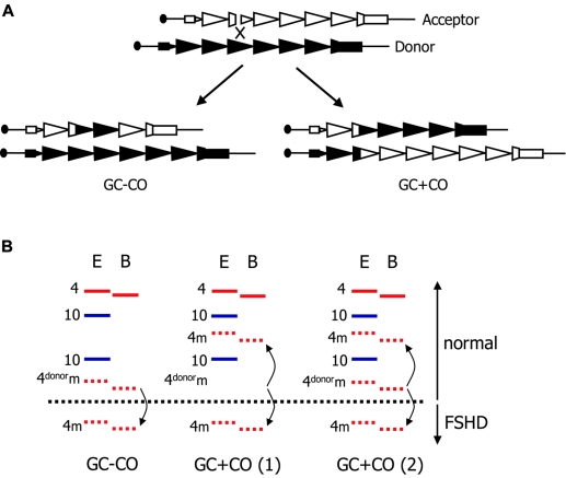
Mechanism of mitotic contractions of D4Z4 in FSHD. A, Generally, repeat rearrangements are induced by double-strand breaks, in this case within D4Z4 (triangles). Break repair through gene conversion with crossover (GC+CO) or without crossover (GC−CO) requires the pairing of the damaged DNA strand (acceptor indicated by unblackened triangles) with the homologous donor (blackened triangles). In FSHD, the sister chromatids are likely the preferred partners in this process. By unequal pairing, the repair of repetitive DNA by GC+CO or by GC−CO is associated with an expansion or contraction of the repeat. B, Schematic representation of somatic mosaicism encountered in FSHD. D4Z4 alleles are visualized in DNA digested by EcoRI (E) and hybridized by probe p13E-11. Chromosome 4 alleles (red) are distinguished from chromosome 10 alleles (blue) by their resistance to BlnI (B). Chromosome 4–derived EcoRI/BlnI fragments are 3 kb smaller because of a single internal BlnI site. GC−CO causes the contraction (or expansion) of a single allele, and the donor alleles remain unchanged. Mosaic (m) alleles are recognized by their reduced signal intensity (dashed lines). During GC+CO, the size of the donor and acceptor alleles are changed. When crossover occurs before the first zygotic division, this yields two cell populations with the newly formed donor and acceptor allele (GC+CO 1). When it occurs in zygotic divisions after the first division, a population of cells, in addition to the two newly formed alleles, will be present carrying the ancestral allele (GC+CO 2).
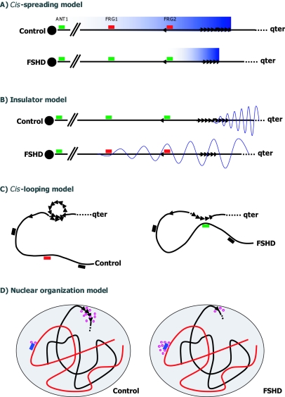
Different models to explain the epigenetic disease mechanism of FSHD. The location of the most well-studied genes (FRG1, FRG2, and ANT1) are indicated. A, In the cis-spreading model, the long D4Z4 repeat (triangles) and nearby sequences share features of heterochromatin. Upon contraction, a local chromatin relaxation (blue gradiation) causes the transcriptional upregulation of 4qter genes (red boxes for downregulated and green boxes for upregulated genes) in a distance-dependent manner, possibly through the action of the D4Z4 repressor complex. B, In the insulator model, D4Z4 acts as a spacer between heterochromatic sequences distal to the D4Z4 repeat and proximal euchromatic sequences. Upon contraction, this insulator function is incomplete, allowing heterochromatinization (wave) of proximal sequencing and transcriptional downregulation of genes within this region. C, The cis-looping model postulates that normally intra-array loops in arrays >11 units prevent the interaction of D4Z4 with genes in cis at a large distance. Disruption of this interaction may cause inappropriate gene expression by long-range interaction with D4Z4. D, Finally, the nuclear organization model predicts that the interaction of 4qter with the nuclear lamina, in which chromatin and transcription factors (purple circles) are tethered, is perturbed in FSHD. This perturbation, in turn, may lead to a misbalance of chromatin and transcription factors at 4qter and unrelated loci.
References
Electronic-Database Information
-
- Online Mendelian Inheritance in Man (OMIM), http://www.ncbi.nlm.nih.gov/Omim/ (for FSHD and ICF)
References
-
- Agresti A, Meneveri R, Siccardi AG, Marozzi A, Corneo G, Gaudi S, Ginelli E (1989) Linkage in human heterochromatin between highly divergent Sau3A repeats and a new family of repeated DNA sequences (HaeIII family). J Mol Biol 205:625–631 - PubMed
-
- Bakker E, Wijmenga C, Vossen RH, Padberg GW, Hewitt J, van der Wielen M, Rasmussen K, Frants RR (1995) The FSHD-linked locus D4F104S1 (p13E-11) on 4q35 has a homologue on 10qter. Muscle Nerve 2:S39–S44 - PubMed
-
- Ballarati L, Piccini I, Carbone L, Archidiacono N, Rollier A, Marozzi A, Meneveri R, Ginelli E (2002) Human genome dispersal and evolution of 4q35 duplications and interspersed LSau repeats. Gene 296:21–27 - PubMed
-
- Coppee F, Matteotti C, Ansseau E, Sauvage S, Leclercq I, Leroy A, Marcowycz A, Gerbaux C, Figlewicz D, Ding H, Belayew A (2004) The DUX gene family and FSHD. In: Upadhyaya M, Cooper DN (eds) Facioscapulohumeral muscular dystrophy: clinical medicine and molecular cell biology. Garland Science, Oxon, pp 117–134
Publication types
MeSH terms
Associated data
- Actions
LinkOut - more resources
Full Text Sources
Other Literature Sources
