. 2005 Feb;86(1):57-66.
doi: 10.1111/j.0959-9673.2005.00414.x.

Histopathology of spleen allograft rejection in miniature swine

Affiliations

Histopathology of spleen allograft rejection in miniature swine

Frank J M F Dor et al. Int J Exp Pathol. 2005 Feb.

Abstract

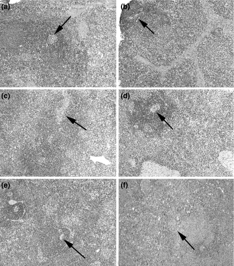
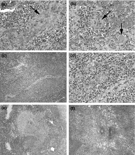
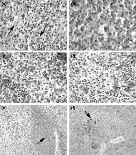
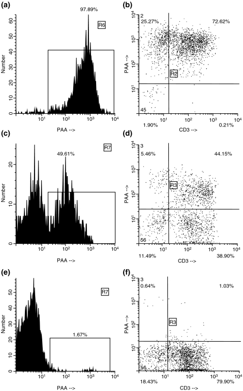
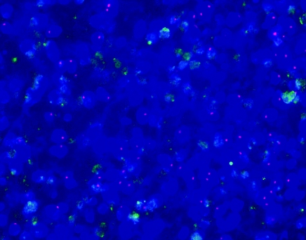
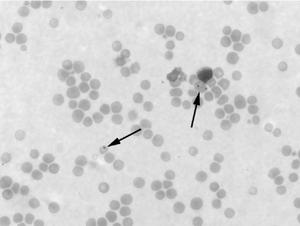
Spleen transplantation (SpTx) has established donor-specific tolerance in rodents, but not in large animals or humans. We report the histopathology of rejection in an established model of SpTx in major histocompatibility complex (MHC)-defined miniature swine. Of the 17 SpTx, rejection was observed in two grafts transplanted into untreated, MHC-matched, minor antigen-disparate recipients (group 1, n=4), but not in the two that received a 12-day course of cyclosporin A (CyA). Rejection also occurred in five grafts transplanted into fully MHC-disparate recipients (group 2, n=12), one of which was untreated and four of which received some form of immunosuppressive therapy. One recipient of an MHC class-I-mismatched spleen treated with 12 days of CyA did not show rejection. Following biopsy and/or necropsy, fixed allograft tissue sections were treated with multiple stains, immunohistochemical markers and TUNEL assay. Common features of rejection occurred in grafts from both groups, but with varying time courses. Necrosis developed as early as day 8 in group 2 and day 27 in group 1, ranging from focal fibrinoid necrosis of arteriolar walls and sinusoids to diffuse liquefactive necrosis, usually associated with haemorrhage. Other features of rejection included white pulp expansion by atypical cells and decreased staining of basement membranes and reticular fibres. A doubling of the baseline TUNEL index preceded histologically identifiable rejection. This study establishes histologic guidelines for diagnosing and, perhaps, in future studies, predicting acute rejection of splenic allografts transplanted across known histocompatibility barriers in a large-animal model.


Figures

Figure 1
Figure 1
Histology of porcine spleen before transplantation of one graft and at progressive time points after transplantation in a graft that was not rejected. Normal features include those of white pulp (arrows mark central arteries in Malpighian corpuscles), red pulp, trabeculae and clusters of histiocytes. (a) Baseline biopsy, before transplantation (pig #14867, group 1) (H&E, ×100); biopsies of graft of pig #15111 (group 1) at (b) 1 h, (c) 15 days, (d) 27 days, (e) 139 days and (f) 362 days after SpTx (H&E, ×100). H&E, hematoxylin and eosin; SpTx, spleen transplantation.
Figure 2
Figure 2
Histology of splenic graft rejection at progressive stages. (a and b) Grade 1 (mild), focal, fibrinoid necrosis of sinusoidal walls (arrow), associated with fibrin thrombi in small vessels (pig #14526, group 1, 57 days after SpTx) (arrows). (c and d) Grade 2 (moderate), marked expansion of PALS around central artery (H&E, ×100), consisting of pleomorphic, atypical lymphoid cells (H&E, ×400) (pig #14867, group 1, 15 days after SpTx). Grade 3 borderline severe (e) and severe (f), focal to diffuse liquefactive necrosis, respectively (pig #14867, group 1, 28 days after SpTx) (H&E, ×100). H&E, hematoxylin and eosin; PALS, periarteriolar lymphatic sheaths; SpTx, spleen transplantation.
Figure 3
Figure 3
Subtle histological changes, which preceded frank rejection. (a and b) The regular network of reticular fibres of the red pulp (arrows) is degraded (b), 4 days after SpTx (pig #14534, group 2) (PAS stain, ×600). The normal (c) and centrally disrupted (d) patterns of reticular fibres are again observed with further staining (pig #14526, group 1, 57 days after SpTx) (reticulin stain, ×600). A TUNEL index (TI) at baseline (e) on the day of SpTx (pig #14867, group 1) is increased more than two-fold (f) 15 days after SpTx, before a subsequent biopsy showed histologic evidence of rejection 28 days after SpTx (arrows mark TUNEL+ cells) (magnification: ×200). PAS, peridodic acid-Schiff; SpTx, spleen transplantation.
Figure 4
Figure 4
Flow cytometry of a donor (PAA+) spleen (donor to pig #15410) before explantation, demonstrating the percentage of PAA+ cells (97.89%) (a) and the profile of CD3+PAA+ cells (b). Flow cytometry of an accepted spleen graft after cessation of CyA (pig #15051, group 2). Replacement with recipient cells is indicated by the presence of PAA– cells. The spleen graft still consists of 49.61% of donor cells (c), but some CD3+PAA– recipient cells can be seen (d). Almost complete repopulation of the spleen graft by recipient cells occurred (pig #15410, group 2, 183 days after SpTx). Only 1.67% of the cells are still of donor origin (e). Virtually, all CD3+ cells (f) are of recipient type (PAA–). CyA, cyclosporin A; PAA, pig allelic antigen; SpTx, spleen transplantation.
Figure 5
Figure 5
Section of the spleen, which was transplanted from a female donor into a male recipient (pig #14840, group 2, 43 days after SpTx). A notable number of recipient-derived cells with Y chromosomes (red signal) have populated the graft at the time of expanded white pulp (day 43) (FISH technique, ×400). FISH, fluorescence in situ hybridization; SpTx, spleen transplantation.
Figure 6
Figure 6
Red blood cells (arrows) contain Howell–Jolly bodies in a peripheral blood smear of a recipient of a rejected (Grade 3) spleen allograft (pig #15311, group 2, 75 days after SpTx, 2 weeks after spleen graft rejection (charcterized by coagulative necrosis) (Wright's stain, ×640). SpTx, spleen transplantation.

References

    1. Bitter-Suermann H. Survival of unmodified spleen allografts in rats. Nature. 1974;247:465–466. - PubMed
    1. Bitter-Suermann H, Herbertson BM. Morphologic features of spleen allografts in rats. Graft rejection, acceptance, or lethal graft-versus-host-disease in the same donor-recipient pairing. Am. J. Pathol. 1975;81:283–294. - PMC - PubMed
    1. Booster MH, Wijnen RM, van Hooff JP, et al. The role of the spleen in pancreas transplantation. Transplantation. 1993;56:1098–1102. - PubMed
    1. Brigden ML, Pattullo AL. Prevention and management of overwhelming postsplenectomy infection – an update. Crit. Care. Med. 1999;27:836–842. - PubMed
    1. Dammin GJ, Wheeler HB, Montague ACW, Dealy JB, Greenberg JB, Moore FD. The splenic allograft: its course in the unmodified and modified canine recipient. Ann. N. Y. Acad. Sci. 1962;99:861–869. - PubMed

Publication types