

Down-regulation of myasthenogenic T cell responses by a dual altered peptide ligand via CD4+CD25+-regulated events leading to apoptosis
- PMID: 15677327
- PMCID: PMC548575
- DOI: 10.1073/pnas.0409549102
Down-regulation of myasthenogenic T cell responses by a dual altered peptide ligand via CD4+CD25+-regulated events leading to apoptosis
Erratum in
- Proc Natl Acad Sci U S A. 2005 Aug 23;102(34):12288
Abstract
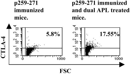
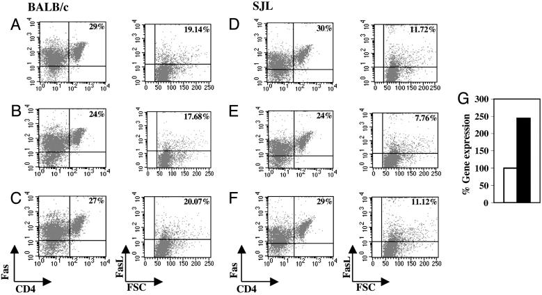
The myasthenogenic peptides p195-212 and p259-271 are sequences of the human acetylcholine receptor and were shown to induce myasthenia gravis-associated immune responses in mice. A dual altered peptide ligand (APL) composed of the two APLs of the myasthenogenic peptides inhibited, in vitro and in vivo, those responses. The aims of this study were to elucidate the events that follow the in vivo treatment with the dual APL and to characterize the cell population that is induced by the latter. We demonstrate here that s.c. administration of the dual APL up-regulates CD4+CD25+ regulatory T cells that are characterized by up-regulated expression of cytotoxic T lymphocyte-associated antigen 4, intracellular and membranal TGF-beta, and Foxp3. Administration of the dual APL to mice concomitant with the immunization with either of the myasthenogenic peptides resulted also in the up-regulation of c-Jun-NH2-terminal kinase activity and of Fas signaling pathway molecules as determined by measuring Fas, Fas ligand, and caspase 8. Thus, our results suggest that the suppression of myasthenia gravis-associated T cell responses exerted by the dual APL is mediated by the CD4+CD25+ immunoregulatory T cell function via TGF-beta or cytotoxic T lymphocyte-associated antigen 4, which further stimulate a cascade of events that up-regulates apoptosis.
Figures


Similar articles
-
Suppression of myasthenogenic responses of a T cell line by a dual altered peptide ligand by induction of CD4+CD25+ regulatory cells.Proc Natl Acad Sci U S A. 2005 Jul 19;102(29):10285-90. doi: 10.1073/pnas.0504578102. Epub 2005 Jul 12. Proc Natl Acad Sci U S A. 2005. PMID: 16014414 Free PMC article.
-
A dual altered peptide ligand down-regulates myasthenogenic T cell responses and reverses experimental autoimmune myasthenia gravis via up-regulation of Fas-FasL-mediated apoptosis.Immunology. 2006 Jul;118(3):413-24. doi: 10.1111/j.1365-2567.2006.02398.x. Immunology. 2006. PMID: 16827902 Free PMC article.
-
A dual altered peptide ligand down-regulates myasthenogenic T cell responses by up-regulating CD25- and CTLA-4-expressing CD4+ T cells.Proc Natl Acad Sci U S A. 2003 May 27;100(11):6676-81. doi: 10.1073/pnas.1131898100. Epub 2003 May 12. Proc Natl Acad Sci U S A. 2003. PMID: 12743364 Free PMC article.
-
Peripheral generation and function of CD4+CD25+ regulatory T cells.Curr Top Microbiol Immunol. 2005;293:115-31. doi: 10.1007/3-540-27702-1_6. Curr Top Microbiol Immunol. 2005. PMID: 15981478 Review.
-
Therapeutic vaccines in autoimmunity.Proc Natl Acad Sci U S A. 2004 Oct 5;101 Suppl 2(Suppl 2):14586-92. doi: 10.1073/pnas.0404826101. Epub 2004 Aug 12. Proc Natl Acad Sci U S A. 2004. PMID: 15308777 Free PMC article. Review.
Cited by
-
The role of CD8+CD28 regulatory cells in suppressing myasthenia gravis-associated responses by a dual altered peptide ligand.Proc Natl Acad Sci U S A. 2007 Oct 30;104(44):17459-64. doi: 10.1073/pnas.0708577104. Epub 2007 Oct 23. Proc Natl Acad Sci U S A. 2007. PMID: 17956982 Free PMC article.
-
Suppression of myasthenogenic responses of a T cell line by a dual altered peptide ligand by induction of CD4+CD25+ regulatory cells.Proc Natl Acad Sci U S A. 2005 Jul 19;102(29):10285-90. doi: 10.1073/pnas.0504578102. Epub 2005 Jul 12. Proc Natl Acad Sci U S A. 2005. PMID: 16014414 Free PMC article.
-
Mucosal administration of an altered CII263-272 peptide inhibits collagen-induced arthritis by suppression of Th1/Th17 cells and expansion of regulatory T cells.Rheumatol Int. 2008 Nov;29(1):9-16. doi: 10.1007/s00296-008-0634-4. Epub 2008 Jul 5. Rheumatol Int. 2008. PMID: 18600328
-
A 50-kDa ERK-like protein is up-regulated by a dual altered peptide ligand that suppresses myasthenia gravis-associated responses.Proc Natl Acad Sci U S A. 2006 Nov 28;103(48):18232-7. doi: 10.1073/pnas.0608896103. Epub 2006 Nov 15. Proc Natl Acad Sci U S A. 2006. PMID: 17108079 Free PMC article.
-
On Peptides and Altered Peptide Ligands: From Origin, Mode of Action and Design to Clinical Application (Immunotherapy).Int Arch Allergy Immunol. 2016;170(4):211-233. doi: 10.1159/000448756. Epub 2016 Sep 20. Int Arch Allergy Immunol. 2016. PMID: 27642756 Free PMC article. Review.
References
Publication types
MeSH terms
Substances
LinkOut - more resources
Full Text Sources
Other Literature Sources
Research Materials
Miscellaneous
