

Extramedullary myeloid tumour (EMMT) of the gallbladder
- PMID: 15677545
- PMCID: PMC1770561
- DOI: 10.1136/jcp.2004.019729
Extramedullary myeloid tumour (EMMT) of the gallbladder
Abstract
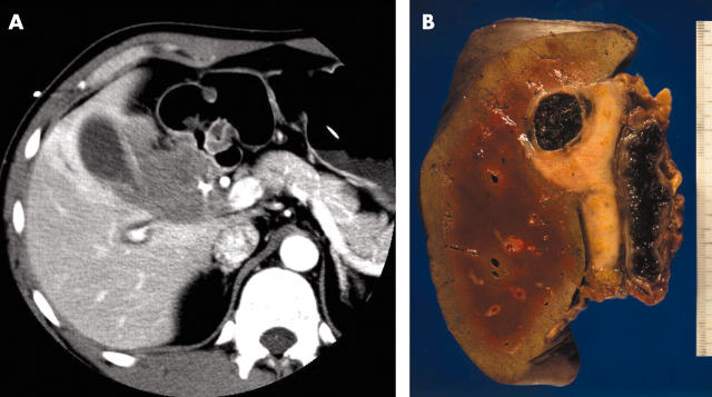
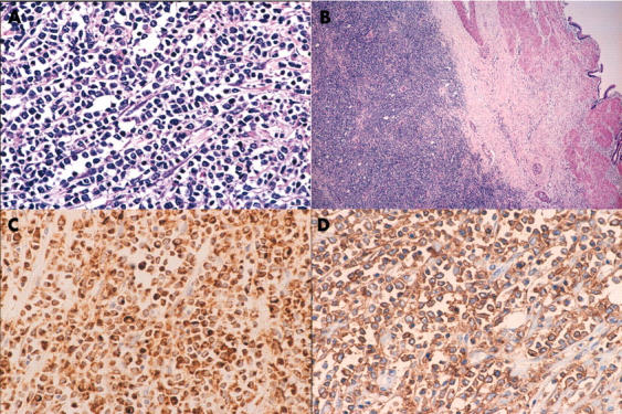
This report describes a rare case of an extramedullary myeloid tumour (EMMT) of the gallbladder in a patient without leukaemia. A 33 year old man visited a local hospital because of jaundice. Abdominal computed tomography revealed a tumorous mass measuring 6.0 x 4.5 cm and involving the entire gallbladder. A percutaneous needle biopsy was attempted, but because adenocarcinoma could not be completely ruled out, the use of undue force was considered dangerous. Under a preoperative diagnosis of gallbladder carcinoma, a hepatopancreatoduodenectomy was performed. The tumour cells exhibited various amounts of eosinophilic cytoplasm, had medium sized round nuclei with indentation and grooving, and were strongly immunoreactive for myeloperoxidase, CD43, and c-kit protein (CD117). After surgery, the patient underwent combination chemotherapy as prescribed for cases of acute myeloblastic leukaemia. The patient did not develop acute leukaemia during a follow up period of four years. In conclusion, a correct diagnosis of EMMT can be made using appropriate immunohistochemical staining.
Figures
Similar articles
-
Disseminated extramedullary myeloid tumor of the gallbladder without involvement of the bone marrow.Am J Hematol. 2007 Jan;82(1):65-8. doi: 10.1002/ajh.20748. Am J Hematol. 2007. PMID: 16947321
-
Myeloid sarcoma of the extrahepatic bile ducts presenting as obstructive jaundice.APMIS. 2006 Sep;114(9):666-8. doi: 10.1111/j.1600-0463.2006.apm_491.x. APMIS. 2006. PMID: 16948823
-
Myeloid sarcoma of the breast in an aleukemic patient: a rare entity in an uncommon location.Malays J Pathol. 2015 Apr;37(1):63-6. Malays J Pathol. 2015. PMID: 25890617
-
State of the art in myeloid sarcoma.Int J Lab Hematol. 2011 Dec;33(6):555-65. doi: 10.1111/j.1751-553X.2011.01361.x. Epub 2011 Aug 24. Int J Lab Hematol. 2011. PMID: 21883967 Review.
-
Neuroendocrine tumor of the gallbladder treated with neoadjuvant chemotherapy and surgery.Am Surg. 2014 Mar;80(3):318-20. Am Surg. 2014. PMID: 24666880 Review. No abstract available.
Cited by
-
An Unexpected Cause of Recurrent Jaundice after Resolution of Acute Hepatitis E.Case Rep Gastroenterol. 2020 Aug 14;14(2):415-419. doi: 10.1159/000508425. eCollection 2020 May-Aug. Case Rep Gastroenterol. 2020. PMID: 32999642 Free PMC article.
-
Acute Myeloid Leukemia in a Cholecystectomy Specimen.Case Rep Pathol. 2022 Sep 26;2022:2956052. doi: 10.1155/2022/2956052. eCollection 2022. Case Rep Pathol. 2022. PMID: 36199751 Free PMC article.
-
Granulocytic sarcoma: An uncommon cause of systemic inflammatory response syndrome.Clin Case Rep. 2019 Jan 31;7(3):469-473. doi: 10.1002/ccr3.1779. eCollection 2019 Mar. Clin Case Rep. 2019. PMID: 30899474 Free PMC article.
-
Myeloid sarcomas: a histologic, immunohistochemical, and cytogenetic study.Diagn Pathol. 2007 Oct 31;2:42. doi: 10.1186/1746-1596-2-42. Diagn Pathol. 2007. PMID: 17974004 Free PMC article.
-
Relapse of acute myeloid leukemia manifested by cholecystitis: A case report and review of the literature.Int J Surg Case Rep. 2014;5(6):302-5. doi: 10.1016/j.ijscr.2014.02.003. Epub 2014 Mar 19. Int J Surg Case Rep. 2014. PMID: 24780774 Free PMC article.
References
-
- Neiman RS, Barcos M, Berard C, et al. Granulocytic sarcoma: a clinicopathologic study of 61 biopsied cases. Cancer 1981;48:1426–37. - PubMed
-
- Menasce LP, Banerjee SS, Beckett E, et al. Extra-medullary myeloid tumor (granulocytic sarcoma) is often misdiagnosed: a study of 26 cases. Histopathology 1999;34:391–8. - PubMed
-
- Hutchison RE, Kurec AS, Davey FR. Granulocytic sarcoma. Clin Lab Med 1990;10:889–901. - PubMed
-
- Jian C, Rudolph R, Yanuck III, et al. c-kit (CD117) reactivity in extramedullary myeloid tumor/granulocytic sarcoma. Arch Pathol Lab Med 2001;125:1448–52. - PubMed
Publication types
MeSH terms
Substances
LinkOut - more resources
Full Text Sources
Medical
Research Materials