. 2005 May 15;105(10):3979-86.
doi: 10.1182/blood-2004-08-3112. Epub 2005 Jan 27.

Expression of the human germinal center-associated lymphoma (HGAL) protein, a new marker of germinal center B-cell derivation

Affiliations

Expression of the human germinal center-associated lymphoma (HGAL) protein, a new marker of germinal center B-cell derivation

Yasodha Natkunam et al. Blood. .

Abstract

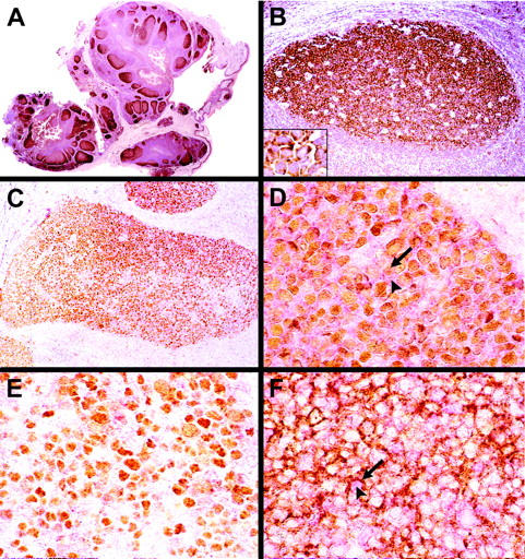
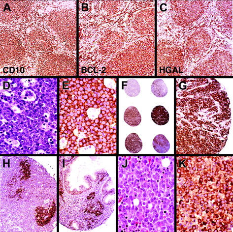
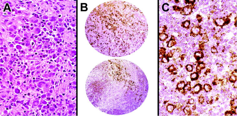
We identified the human germinal center-associated lymphoma (HGAL) in gene-expression profiling studies of diffuse large B-cell lymphoma (DLBCL). The expression of HGAL correlated with survival in patients with DLBCL. The HGAL gene is the human homolog of M17, a mouse gene expressed specifically in normal germinal center (GC) B cells. We generated a monoclonal antibody against the HGAL protein and show that HGAL is expressed in the cytoplasm of GC lymphocytes and in lymphomas of GC derivation. Among 727 lymphomas tested by immunohistochemistry on tissue microarrays, HGAL staining was found in follicular lymphomas (103 of 107), Burkitt lymphomas (40 of 40), mediastinal large B lymphomas (7 of 8), and in DLBCLs (103 of 151). Most marginal zone lymphomas lacked HGAL staining. Lymphocyte-predominant Hodgkin lymphomas (12 of 17) and, surprisingly, classical Hodgkin lymphomas (78 of 107) were found to be positive. Hierarchical clustering of comparative immunohistologic results in DLBCLs demonstrates that the expression of HGAL is similar to 2 other GC-associated proteins, BCL6 and CD10, but different from 2 markers associated with a non-GC phenotype, MUM1/IRF4 and BCL2. The restricted expression and GC specificity of HGAL protein suggest that it may have an important role in the diagnosis of specific lymphomas, and, potentially in the identification of subtypes associated with different prognoses.


Figures

Figure 1.
Figure 1.
Localization of HGAL protein and specificity of anti-HGAL monoclonal antibody. (A) Confocal immunofluorescence microscopy using a FITC-conjugated anti-V5 antibody in transfected HeLa cells shows that the HGAL protein is localized to the cytoplasm (green fluorescence). The nuclei are counterstained with an orange Hoechst dye (original magnification, ×1000). (B) Western blot analysis using the anti-HGAL monoclonal antibody shows a specific band corresponding to HGAL protein expression in Raji cells (positive control) and in HeLa cells stably transfected with pcDNA3.1 HGAL-V5 but not in native or mock-transfected HeLa cells.
Figure 2.
Figure 2.
Correlation between HGAL mRNA and protein expression. HGAL mRNA and protein expression were assessed by real-time RT-PCR and Western blot using the anti-HGAL antibody, respectively, in GC-like (SU-DHL-4, OCI-LY7) and in non–GC-like (RCK8, OCI-LY3, OCI-LY10) DLBCL cell lines and in the Jurkat T-cell line. Actin was used as a loading control.
Figure 3.
Figure 3.
Immunohistochemical staining with anti-HGAL monoclonal antibody in tonsil tissues. (A-B) GCs are highlighted in a normal tonsil; an increased intensity of staining is seen within cells of the proliferating pole or dark zone of GCs, whereas mantle and marginal zones lack staining. Inset shows a higher magnification of HGAL staining in germinal-center cells. (C) Double immunohistologic labeling for HGAL (red cytoplasmic stain) and BCL6 (brown nuclear stain) are colocalized in a significant number of cells within the GC. (D) A subset of GC cells shows coexpression of BCL6 (arrow) and HGAL (arrowhead). (E) A subset of GC cells located in the proliferative pole of the GC shows staining for BCL6 but not for HGAL. (F) Double immunohistologic labeling for HGAL (red cytoplasmic stain, arrowhead) and CD10 (brown membrane stain, arrow) show that a majority of GC cells coexpress these 2 proteins although cells expressing only HGAL or CD10 are also present. Original magnifications: A, ×30; B and C, ×150; D-F and B inset, ×600.
Figure 4.
Figure 4.
Immunohistochemical staining with anti-HGAL monoclonal antibody in lymphomas. (A-C) Follicular lymphoma stained with the CD10, BCL2, and anti-HGAL monoclonal antibody highlights neoplastic follicles and interfollicular B cells. (D) Hematoxylin and eosin-stained section of Burkitt lymphoma shows uniform cells admixed with starry sky histiocytes. (E) Burkitt lymphoma cells show staining for HGAL protein. (F) TMA cores of DLBCL show a range of staining from none to weak/partial staining to intense staining of all neoplastic cells. (G) High magnification of a core of a DLBCL shows moderate to strong staining within the cytoplasm. (H-I) Extranodal marginal zone B-cell lymphoma of the lymph node and gastrointestinal tract lacks staining for HGAL although the stain highlights entrapped GCs within the neoplastic infiltrate. (J) Hematoxylin and eosin–stained section of plasma cell myeloma shows sheets of atypical plasma cells with prominent nucleoli. (K) Plasma cell myeloma shows staining for HGAL protein. Original magnifications: A-C, H, and I, ×150; F, ×30; G, ×300; D, E, J, and K, ×600.
Figure 5.
Figure 5.
HGAL staining in Hodgkin lymphoma. Immunohistochemical staining for HGAL protein highlights large atypical cells in a case of nodular sclerosis Hodgkin lymphoma on 2.0-mm cores on a TMA. The staining is localized within the cytoplasm of Hodgkin cells. Original magnifications: A and C, ×30; B, ×600.
Figure 6.
Figure 6.
Hierarchical clustering of protein expression data. The expression patterns of 5 proteins, HGAL, CD10, BCL6, MUM1/IRF4 (MUM1), and BCL2, predicted to be associated with distinct subtypes of DLBCL based on the cell of origin by gene expression profiling studies are shown. Positive staining is indicated as red and the lack of staining as green. Stains that were not interpretable are indicated in white. The branching pattern of the dendrogram reflects similarities in the patterns of reactivity of the antibodies, with short branches denoting a high degree of similarity in expression pattern of the cognate protein across 151 DLBCL cases. HGAL clusters with BCL6 and CD10, associated with a GC phenotype, on one branch of the dendrogram (indicated in orange), whereas MUM1 and BCL2, associated with an ABL phenotype, cluster on a separate branch (indicated in blue).

Similar articles

Cited by

References

    1. Alizadeh AA, Eisen MB, Davis RE, et al. Distinct types of diffuse large B-cell lymphoma identified by gene expression profiling. Nature. 2000;403: 503-511. - PubMed
    1. Rosenwald A, Wright G, Chan WC, et al. The use of molecular profiling to predict survival after chemotherapy for diffuse large-B-cell lymphoma. N Engl J Med. 2002;346: 1937-1947. - PubMed
    1. Shipp MA, Ross KN, Tamayo P, et al. Diffuse large B-cell lymphoma outcome prediction by gene-expression profiling and supervised machine learning. Nat Med. 2002;8: 68-74. - PubMed
    1. Lossos IS, Alizadeh AA, Rajapaksa R, Tibshirani R, Levy R. HGAL is a novel interleukin-4-inducible gene that strongly predicts survival in diffuse large B-cell lymphoma. Blood. 2003;101: 433-440. - PubMed
    1. Pan Z, Shen Y, Du C, et al. Two newly characterized germinal center B-cell-associated genes, GCET1 and GCET2, have differential expression in normal and neoplastic B cells. Am J Pathol. 2003;163: 135-144. - PMC - PubMed

Publication types

MeSH terms