. 2005 Feb 8;102(6):2069-74.
doi: 10.1073/pnas.0409736102. Epub 2005 Jan 27.

Overexpression of PCSK9 accelerates the degradation of the LDLR in a post-endoplasmic reticulum compartment

Affiliations

Overexpression of PCSK9 accelerates the degradation of the LDLR in a post-endoplasmic reticulum compartment

Kara N Maxwell et al. Proc Natl Acad Sci U S A. .

Abstract

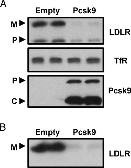
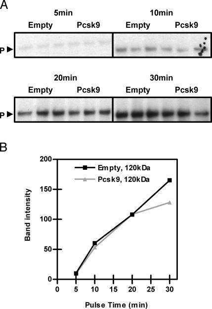
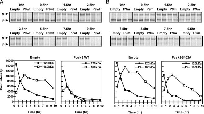
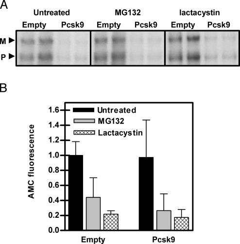
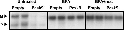
Proprotein convertase subtilisin kexin 9 (PCSK9) is a member of the subtilisin serine protease family with an important role in cholesterol metabolism. PCSK9 expression is regulated by dietary cholesterol in mice and cellular sterol levels in cell culture via the sterol regulatory element binding protein transcription factors, and mutations in PCSK9 are associated with a form of autosomal dominant hypercholesterolemia. Overexpression of PCSK9 in mice leads to increased total and low-density lipoprotein (LDL) cholesterol levels because of a decrease in hepatic LDL receptor (LDLR) protein with normal mRNA levels. To study the mechanism, PCSK9 was overexpressed in human hepatoma cells, HepG2, by adenovirus. Overexpression of PCSK9 in HepG2 cells caused a decrease in whole-cell and cell-surface LDLR levels. PCSK9 overexpression had no effect on LDLR synthesis but caused a dramatic increase in the degradation of the mature LDLR and a lesser increase in the degradation of the precursor LDLR. In contrast, overexpression of a catalytically inactive mutant PCSK9 prevented the degradation of the mature LDLR; whereas increased degradation of the precursor LDLR still occurred. The PCSK9-induced degradation of the LDLR was not affected by inhibitors of the proteasome, lysosomal cysteine proteases, aspartic acid proteases, or metalloproteases. The PCSK9-induced degradation of the LDLR was shown to require transport out of the endoplasmic reticulum. These results indicate that overexpression of PCSK9 induces the degradation of the LDLR by a nonproteasomal mechanism in a post-endoplasmic reticulum compartment.


Figures

Fig. 1.
Fig. 1.
Overexpression of PCSK9 in HepG2 cells decreased whole-cell and cell-surface LDLR. (A) HepG2 cells were infected with control adenovirus (Empty) or PCSK9 adenovirus (Pcsk9), and whole-cell lysates were collected after 36 h. Cell lysates were subjected to Western blotting for LDLR, transferrin receptor (TfR), and PCSK9. M, mature LDLR; P, precursor LDLR; P, pro-PCSK9; C, cleaved PCSK9. (B) HepG2 cells were infected with empty adenovirus or PCSK9 adenovirus. After 36 h, cell-surface proteins were biotinylated at 4°C and lysates were collected and immunoprecipitated for the LDLR. Proteins were visualized with avidin-HRP. The results presented in this and other figures are representative of at least three experiments each.
Fig. 2.
Fig. 2.
Overexpression of PCSK9 did not affect synthesis of the LDLR. (A) HepG2 cells were infected with empty or PCSK9 adenovirus (Pcsk9). Cells were pulse-labeled with 35S-Met/Cys for 5, 10, 20, and 30 min and immunoprecipitated for the LDLR. P, precursor LDLR. (B) Quantification of the results in A; band intensities were quantified with image pro plus.
Fig. 3.
Fig. 3.
Overexpression of PCSK9 accelerated the degradation of the mature LDLR in a catalytically dependent mechanism. (A) HepG2 cells were infected with empty or PCSK9 adenovirus (Pcsk9WT or P9wt). (B) HepG2 cells were infected with empty or PCSK9S402A adenovirus (Pcsk9S402A or P9m). Cells were pulse labeled with 35S-Met/Cys for 30 min, chased for 0, 0.5, 1.5, 2.5, 3.5, 5.5, 7.5, and 9.5 h, and immunoprecipitated for the LDLR. Band intensities were measured by using imagequant and graphed below. M, mature LDLR; P, precursor LDLR.
Fig. 4.
Fig. 4.
PCSK9-induced degradation of the LDLR did not depend on the proteasome. (A) HepG2 cells were infected with empty or PCSK9 adenovirus (Pcsk9). Cells were pulse-labeled with 35S-Met/Cys for 30 min, chased for 4 h in the presence of the proteasome inhibitors MG132 and lactacystin, and immunoprecipitated for the LDLR. M, mature LDLR; P, precursor LDLR. (B) To measure proteasome activity in these cells, lysates were incubated with the proteasome substrate Suc-LLVY-AMC for 1 h. Fluorescence due to cleaved AMC by the proteasome was measured and compared to background fluorescence.
Fig. 5.
Fig. 5.
PCSK9-induced degradation of the LDLR depends on exit from the ER. HepG2 cells were infected with empty or PCSK9 adenovirus (Pcsk9). Cells were pulse-labeled with 35S-Met/Cys for 30 min, chased for 4 h in the presence of BFA and BFA plus nocodazole (noc), and immunoprecipitated for the LDLR. M, mature LDLR; P, precursor LDLR.

Similar articles

Cited by

References

    1. Bergeron, F., Leduc, R. & Day, R. (2000) J. Mol. Endocrinol. 24, 1–22. - PubMed
    1. Gensberg, K., Jan, S. & Matthews, G. M. (1998) Semin. Cell Dev. Biol. 9, 11–17. - PubMed
    1. Seidah, N. G. & Chretien, M. (1999) Brain Res. 848, 45–62. - PubMed
    1. Taylor, N. A., Van De Ven, W. J. & Creemers, J. W. (2003) FASEB J. 17, 1215–1227. - PubMed
    1. Zhou, A., Webb, G., Zhu, X. & Steiner, D. F. (1999) J. Biol. Chem. 274, 20745–20748. - PubMed

Publication types