. 2005 Feb 23;24(4):753-65.
doi: 10.1038/sj.emboj.7600569. Epub 2005 Jan 27.

Plk1 docking to GRASP65 phosphorylated by Cdk1 suggests a mechanism for Golgi checkpoint signalling

Affiliations

Plk1 docking to GRASP65 phosphorylated by Cdk1 suggests a mechanism for Golgi checkpoint signalling

Christian Preisinger et al. EMBO J. .

Abstract

GRASP65, a structural protein of the Golgi apparatus, has been linked to the sensing of Golgi structure and the integration of this information with the control of mitotic entry in the form of a Golgi checkpoint. We show that Cdk1-cyclin B is the major kinase phosphorylating GRASP65 in mitosis, and that phosphorylated GRASP65 interacts with the polo box domain of the polo-like kinase Plk1. GRASP65 is phosphorylated in its C-terminal domain at four consensus sites by Cdk1-cyclin B, and mutation of these residues to alanine essentially abolishes both mitotic phosphorylation and Plk1 binding. Expression of the wild-type GRASP65 C-terminus but not the phosphorylation defective mutant in normal rat kidney cells causes a delay but not the block in mitotic entry expected if this were a true cell cycle checkpoint. These findings identify a Plk1-dependent signalling mechanism potentially linking Golgi structure and cell cycle control, but suggest that this may not be a cell cycle checkpoint in the classical sense.


Figures

Figure 1
Figure 1
Cdk1–cyclin B is the major GRASP65 kinase in the mitotic HeLa extract. (A) Mitotic HeLa extract was fractionated over HiTRAP-Q ion exchange columns as described in Materials and methods. (B) The QI peak of kinase activity was further fractionated over heparin sepharose to yield a single peak of activity. (C, D) The HI peak was then separated by size exclusion chromatography on a Superose 6 HR10/30 column. This resolved three peaks of GRASP65 kinase activity. Molecular size standards are indicated along the top of the panels. (E) QII, the second peak from the initial ion exchange column, was separated by size exclusion chromatography on a Superose 6 HR10/30 column. This indicates that the kinase activity is associated with a protein of apparent molecular size 30 kDa, possibly Cdk1 free of cyclin B. The load (L) and the numbered fractions were tested for kinase activity towards GRASP65 and GRASP55, and Western blotted for Plk1, Cdk1, cyclin B1, and Mps1 as indicated.
Figure 2
Figure 2
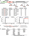
Mapping of phosphorylation sites in GRASP65. (A) Schematic of GRASP65 indicating the two N-terminal PDZ-like domains and GM130 binding region. Phosphorylation sites are shown with the phosphorylated residues numbered and marked in red. The table summarises data on the kinases that phosphorylate these sites and whether or not they are modified in interphase or mitosis. Phosphorylation of S372 was observed under some conditions, but this was substoichiometric and could not be assigned to any kinase with confidence, although it does fall within a Plk1 consensus sequence, and is thus omitted from the table. (B) MALDI-TOF spectra showing a peptide corresponding to GRASP65 amino acids 359–388, phosphorylated with SI and SII from the Superose 6 fractionation of mitotic extract (see Figure 1C). A peak corresponding to the addition of a single phosphate is observed with the Cdk1–cyclin B-containing SII fraction. (C) Fragmentation of the phosphorylated GRASP65359−388 peptide by tandem mass spectrometry gives daughter ions derived from the N- and C-termini of the peptide indicating that serine 376 (pS), shown in red, is the phosphorylated residue. (D) Phosphorylation sites predicted to be equivalent to those identified in rat GRASP65 are shown for mouse, human, and chicken GRASP65.
Figure 3
Figure 3
Cdk1–cyclin B is the major GRASP65 kinase in mitotic HeLa extracts. (A) GRASP65 and the GRASP65 8A mutant where the following residues have been mutated to alanine (T220, S216, 217, 277, 367, 372, 376, and 400) were incubated with buffer (control), and interphase and mitotic cytosols. Substrate loading was controlled by Western blotting for the HA epitope tag present on the recombinant GRASP65, and an autoradiograph indicates the amount of [γ-32P] incorporation. (B) GRASP65, GRASP65 8A, and the GRASP65 4A mutants where the following residues have been mutated to alanine (T220, S217, 277, and 376) were incubated with buffer (−), or in the presence (+) of Cdk1–cyclin B. Substrate loading was controlled by Coomassie brilliant blue (CBB) staining, and an autoradiograph indicates the amount of [γ-32P] incorporation. (C) Phospho-antibodies pT220, pS367, and pS376 were used to Western blot recombinant HA-tagged GRASP65 treated with buffer, and interphase and mitotic cytosols. Control experiments were performed with antibodies preincubated with the phosphopeptide antigen to compete specific antibody reactivity (+peptide), and GRASP65 phosphorylated with mitotic cytosol was treated with or without alkaline phosphatase. GRASP65 loading was controlled with the HA epitope antibody.
Figure 4
Figure 4
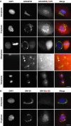
GRASP65 pS376 phosphorylation correlates with entry into mitosis. NRK cells were stained with DAPI to reveal DNA (blue) and antibodies to either (A) GRASP65 (green) and GRASP65 pS376 (red), or (B) GM130 (green) and GM130 phosphoserine 25, pS25 (red). Interphase cells and cells at various stages in mitosis are shown. A 4.6-fold enlargement of a region of the metaphase cells shows the presence of GRASP65 and GRASP65 pS376 staining on mitotic Golgi fragments (arrowhead). The bar indicates 10 μm.
Figure 5
Figure 5
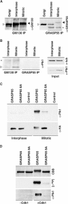
Plk1 interacts with GRASP65 in mitosis. (A) Interphase and mitotic GRASP65–GM130 (GM130 IP) and Golgin45–GRASP55 (GRASP55 IP) complexes were immune precipitated and Western blotted for GM130 and GRASP55, respectively. The asterisk marks a crossreaction with the antibody heavy chain in the GRASP55 blot. (B) Western blot of the GRASP65 and GRASP55 complexes for Plk1. Loading controls for extracts blotted for α-tubulin (α-tub) and Plk1 are shown on the right. Ligand blots were carried out with Plk1 amino acids 305–603 as the probe, and (C) interphase and mitotically or (D) Cdk1–cyclin B phosphorylated GRASP65 and the GRASP65 8A phosphorylation site mutants as the targets.
Figure 6
Figure 6
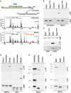
Docking of Plk1 to mitotically phosphorylated cis-Golgi proteins. (A) Schematic of Rab1 indicating the GTP binding domain (green) and prenylation sites. The variable C-terminal region with the phosphorylation site is shown with the phosphorylated residue numbered and marked in red. MALDI-TOF spectra showing a peptide corresponding to Rab1 amino acids 187–205 (m/z 2035.118), after treatment under interphase and mitotic conditions. A peak (m/z 2115.105) corresponding to the addition of a single phosphate is only observed under mitotic conditions. (B) Ligand blots were carried out with Plk1 amino acids 305–603 as the probe, and GST-tagged Rab1 or Rab1 T195A incubated with interphase or mitotic cytosol as the targets. (C) GST-tagged Rab1 or Rab1 T195A were incubated with buffer (−) or Cdk1–cyclin B (+). Samples were analysed by autoradiography ([γ-32P]) or ligand blotting with Plk1. Ligand blots were carried out with Plk1 amino acids 305 to 603 as the probe, and (D) MBP-tagged GM1301−271 or Rab1, (E) HA-tagged GRASP65 or GRASP55, and (F) GST-tagged Rab1 or Rab2 incubated with interphase or mitotic cytosol as the targets.
Figure 7
Figure 7
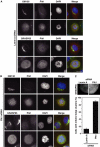
Effect of Plk1 depletion on mitotic Golgi fragmentation. HeLa cells or HeLa cells stably expressing GRASP65-GFP were treated with siRNA duplexes for (A) lamin A and (B) Plk1 for 72 h, and then stained with antibodies to GM130 (green) and Plk1 (red), or stained with an antibody to Plk1 (red) and GRASP65 visualised using GFP (green). DNA was stained with DAPI (blue). Bar indicates 10 μm. (C) Enlarged images showing the increased number of persistent Golgi clusters, marked by GRASP65-GFP, observed in Plk1 siRNA cells. Numbers of mitotic cells containing Golgi clusters were counted for lamin A and Plk1 siRNA and plotted as a bar graph (300 cells per experiment, n=3).
Figure 8
Figure 8
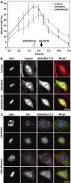
Effect of phosphorylation mutant GRASP65 on mitotic entry. NRK cells expressing the C-terminal domain of wild-type GRASP65 and the phosphorylation defective mutant GRASP65 8A were treated with aphidicolin and then allowed to synchronously passage through mitosis. (A) The mitotic index was counted at the times indicated in the figure after removal of aphidicolin; an average of 150 cells were counted per condition, for three or more experiments. (B, C) The GRASP65 C-terminal domain was visualised by GFP fluorescence, and cells were counterstained with antibodies to α-tubulin or Plk1 (red). DNA was stained with DAPI (blue). Bar indicates 10 μm.

Similar articles

Cited by

References

    1. Acharya U, Mallabiabarrena A, Acharya JK, Malhotra V (1998) Signaling via mitogen-activated protein kinase kinase (MEK1) is required for Golgi fragmentation during mitosis. Cell 92: 183–192 - PubMed
    1. Allan BB, Moyer BD, Balch WE (2000) Rab1 recruitment of p115 into a cis-SNARE complex: programming budding COPII vesicles for fusion. Science 289: 444–448 - PubMed
    1. Altan-Bonnet N, Phair RD, Polishchuk RS, Weigert R, Lippincott-Schwartz J (2003) A role for Arf1 in mitotic Golgi disassembly, chromosome segregation, and cytokinesis. Proc Natl Acad Sci USA 100: 13314–13319 - PMC - PubMed
    1. Bailly E, McCaffrey M, Touchot N, Zahraoui A, Goud B, Bornens M (1991) Phosphorylation of two small GTP-binding proteins of the Rab family by p34cdc2. Nature 350: 715–718 - PubMed
    1. Barr FA, Nakamura N, Warren G (1998) Mapping the interaction between GRASP65 and GM130, components of a protein complex involved in the stacking of Golgi cisternae. EMBO J 17: 3258–3268 - PMC - PubMed

Publication types

MeSH terms