

Brief breath holding may confound functional magnetic resonance imaging studies
- PMID: 15678482
- PMCID: PMC6871742
- DOI: 10.1002/hbm.20086
Brief breath holding may confound functional magnetic resonance imaging studies
Abstract
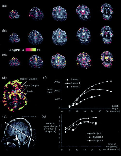
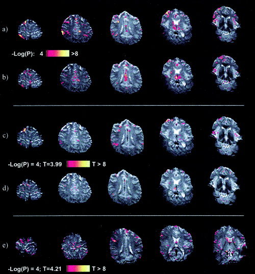
We demonstrate that breath holding of short durations may confound functional magnetic resonance imaging (fMRI) studies. Some subjects may hold their breath for a short time during task performance, especially if the task is challenging. Breath holding may therefore need to be considered specifically when interpreting fMRI experiments. We studied the temporal and spatial characteristics of cerebral T2*-weighted signal during short periods of breath holding by seven individuals in a 3-tesla MR scanner. We demonstrate that breath-holds as short as 3 s can result in regions of significant cerebral activation. More interestingly, we show that focal activation remains present when the data is analysed in a number of different ways, including analyses that correct for motion and model the task epoch as if it were 10 times longer than the actual breath-hold length. These findings have potential relevance for many researchers carrying out fMRI studies.
(c) 2005 Wiley-Liss, Inc.
Figures
References
-
- Abbott D, Jackson G (2001): iBrain–software for analysis and visualisation of functional MR images. Neuroimage 13(Suppl.): 59.
-
- Fox IJ, Crowley WP Jr, Grace JB, Wood EH (1966): Effects of the Valsalva maneuver on blood flow in the thoracic aorta in man. J Appl Physiol 21: 1553–1560. - PubMed
-
- Frackowiak RSJ, Friston KJ, Frith CD, Dolan RJ, Mazziotta JC (1997): Human brain function. San Diego: Academic Press; 528 p.
-
- Friston KJ, Fletcher P, Josephs O, Holmes A, Rugg MD, Turner R (1998): Event‐related fMRI: characterizing differential responses. Neuroimage 7: 30–40. - PubMed
-
- Kastrup A, Kruger G, Glover GH, Neumann‐Haefelin T, Moseley ME (1999): Regional variability of cerebral blood oxygenation response to hypercapnia. Neuroimage 10: 675–681. - PubMed
Publication types
MeSH terms
LinkOut - more resources
Full Text Sources
Medical
