

Anatomics: the intersection of anatomy and bioinformatics
- PMID: 15679867
- PMCID: PMC1571451
- DOI: 10.1111/j.0021-8782.2005.00376.x
Anatomics: the intersection of anatomy and bioinformatics
Abstract
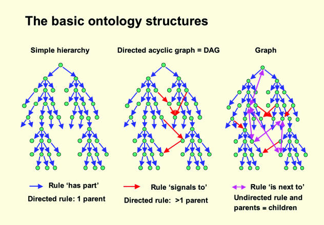
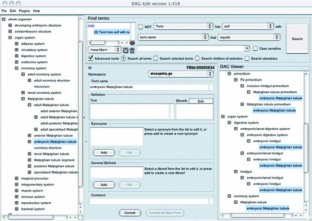
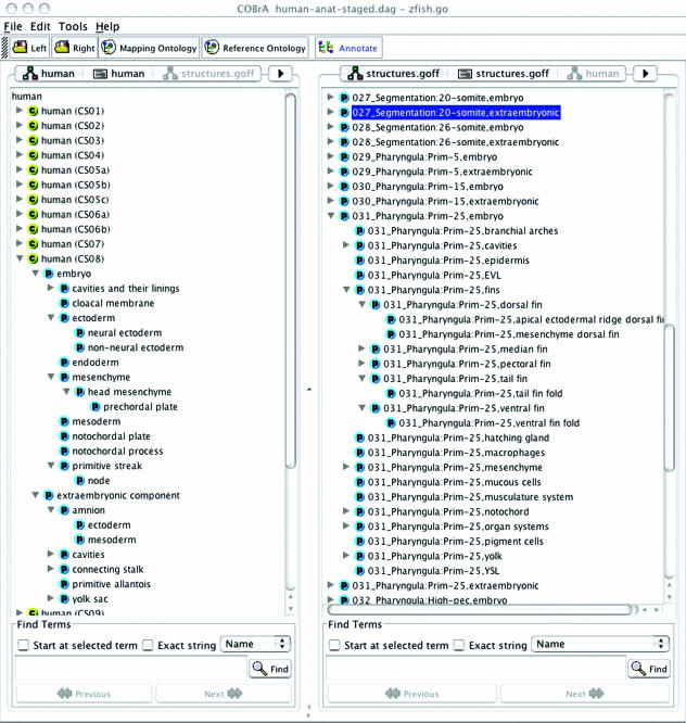
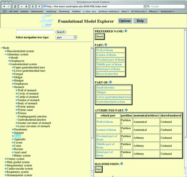
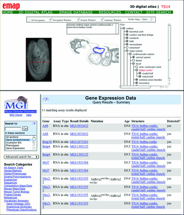
Computational resources are now using the tissue names of the major model organisms so that tissue-associated data can be archived in and retrieved from databases on the basis of developing and adult anatomy. For this to be done, the set of tissues in that organism (its anatome) has to be organized in a way that is computer-comprehensible. Indeed, such formalization is a necessary part of what is becoming known as systems biology, in which explanations of high-level biological phenomena are not only sought in terms of lower-level events, but are articulated within a computational framework. Lists of tissue names alone, however, turn out to be inadequate for this formalization because tissue organization is essentially hierarchical and thus cannot easily be put into tables, the natural format of relational databases. The solution now adopted is to organize the anatomy of each organism as a hierarchy of tissue names and linking relationships (e.g. the tibia is PART OF the leg, the tibia IS-A bone) within what are known as ontologies. In these, a unique ID is assigned to each tissue and this can be used within, for example, gene-expression databases to link data to tissue organization, and also used to query other data sources (interoperability), while inferences about the anatomy can be made within the ontology on the basis of the relationships. There are now about 15 such anatomical ontologies, many of which are linked to organism databases; these ontologies are now publicly available at the Open Biological Ontologies website (http://obo.sourceforge.net) from where they can be freely downloaded and viewed using standard tools. This review considers how anatomy is formalized within ontologies, together with the problems that have had to be solved for this to be done. It is suggested that the appropriate term for the analysis, computer formulation and use of the anatome is anatomics.
Figures
References
-
- Aitken JS, Webber BL, Bard JBL. Part-of relations in anatomy ontologies: a proposal for RDFS and OWL formalisations. Pacific Symp. Bioinformatics. 2004;9:166–177. - PubMed
-
- Baader F, Horrocks I, Sattler U. Description logics. In: Staab S, Studer R, editors. Handbook on Ontologies. Berlin: Springer; 2004. pp. 3–28.
-
- Bard JBL, Rhee SY. Ontologies in biology: design, applications, and future challenges. Nature Rev. Gen. 2004;5:213–222. - PubMed
-
- Birk OS, Casiano DE, Wassif CA, et al. The LIM homeobox gene Lhx9 is essential for mouse gonad formation. Nature. 2000;403:909–913. - PubMed
Publication types
MeSH terms
LinkOut - more resources
Full Text Sources
Medical
