

Development of the pelvis and posterior part of the vertebral column in the Anura
- PMID: 15679868
- PMCID: PMC1571457
- DOI: 10.1111/j.0021-8782.2005.00366.x
Development of the pelvis and posterior part of the vertebral column in the Anura
Abstract
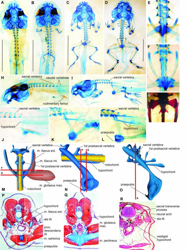
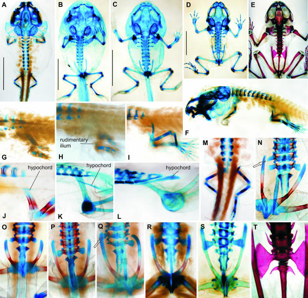
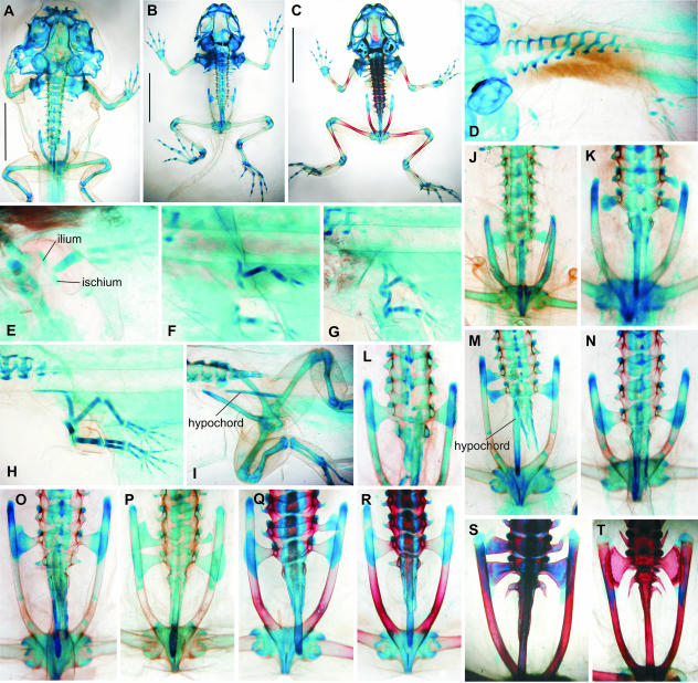
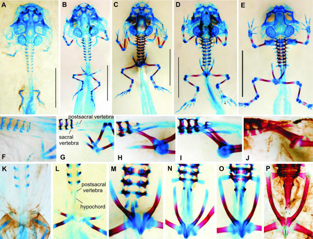
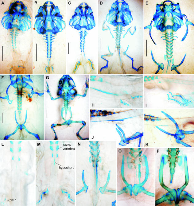
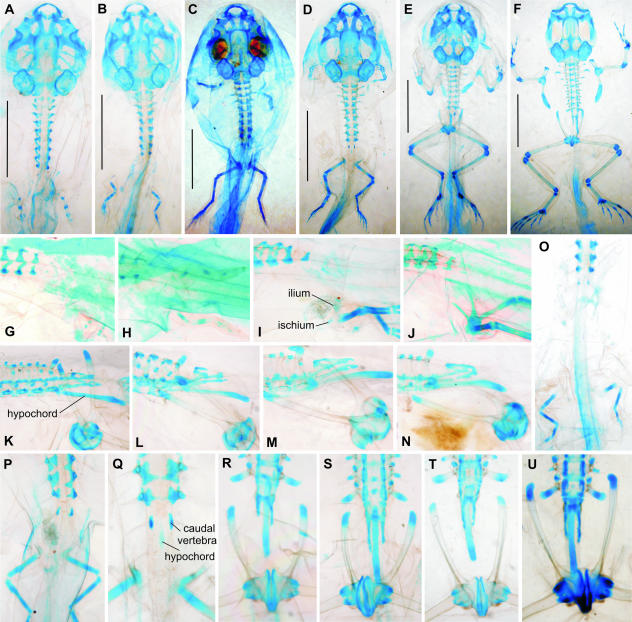
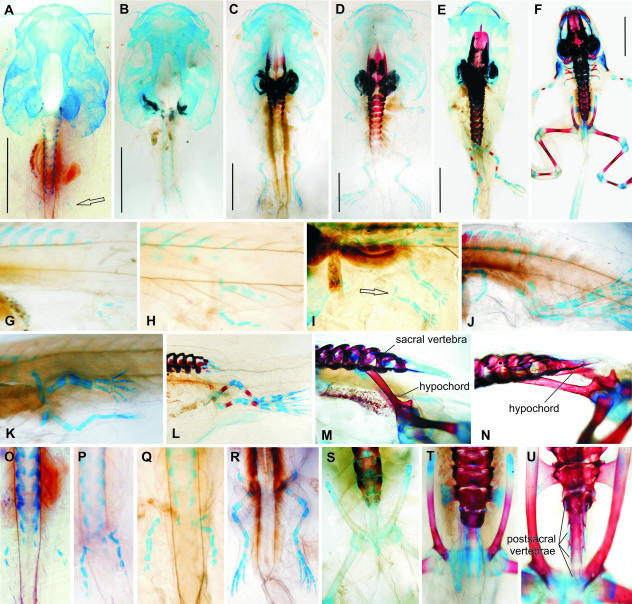
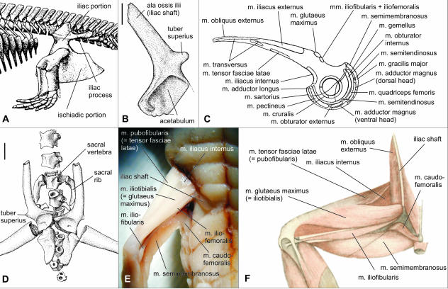
The anuran pelvic girdle is unique among all amphibians in that its acetabular portion is located far posterior to the sacrum, lateral to the postsacral (= caudal) vertebral column, which is reduced to a single rod-like element called the urostyle. This situation in the adult is strikingly different not only from that in ancestral temnospondyls but also in other modern amphibians. Because there is no fossil that would document this evolutionary anatomical modification except for Triadobatrachus, the only data may be inferred from development in modern anurans. We chose seven anuran species (belonging to the genera Discoglossus, Bombina, Pelobates, Bufo, Rana and Xenopus), representing the principal locomotory types (saltation, swimming, crawling and burrowing). Development of the pelvic girdle was studied on cleared and stained whole mounts and partly on serial histological sections. The basic developmental pattern was similar in all species: the pelvis on both sides develops from two centres (puboischiadic and iliac, respectively). The ilium then extends vertically towards the sacral vertebra and later rotates posteriorly so that ultimately the acetabulum is lateral to the tail (= urostyle). Only minor deviations from this pattern were found, mainly associated with differences in water and terrestrial dwelling.
Figures
Similar articles
-
Transformation of the pectoral girdle in the evolutionary origin of frogs: insights from the primitive anuran Discoglossus.J Anat. 2006 Jul;209(1):1-11. doi: 10.1111/j.1469-7580.2006.00583.x. J Anat. 2006. PMID: 16822264 Free PMC article.
-
The evolution of jumping in frogs: morphological evidence for the basal anuran locomotor condition and the radiation of locomotor systems in crown group anurans.J Morphol. 2011 Feb;272(2):149-68. doi: 10.1002/jmor.10902. Epub 2010 Nov 8. J Morphol. 2011. PMID: 21210487
-
Bony-tailed tadpoles: the development of supernumerary caudal vertebrae in larval megophryids (Anura).Evol Dev. 2007 Mar-Apr;9(2):190-202. doi: 10.1111/j.1525-142X.2007.00149.x. Evol Dev. 2007. PMID: 17371401
-
The anuran Bauplan: a review of the adaptive, developmental, and genetic underpinnings of frog and tadpole morphology.Biol Rev Camb Philos Soc. 2007 Feb;82(1):1-25. doi: 10.1111/j.1469-185X.2006.00001.x. Biol Rev Camb Philos Soc. 2007. PMID: 17313522 Review.
-
The natural history of human gait and posture. Part 1. Spine and pelvis.Gait Posture. 2005 Jan;21(1):95-112. doi: 10.1016/j.gaitpost.2004.01.001. Gait Posture. 2005. PMID: 15536039 Review.
Cited by
-
Perichordal Vertebral Column Formation in Rana kobai.J Morphol. 2025 Apr;286(4):e70044. doi: 10.1002/jmor.70044. J Morphol. 2025. PMID: 40181708 Free PMC article.
-
Pelvic and thigh musculature in frogs (Anura) and origin of anuran jumping locomotion.J Anat. 2009 Jan;214(1):100-39. doi: 10.1111/j.1469-7580.2008.01006.x. J Anat. 2009. PMID: 19166476 Free PMC article.
-
Digital dissection of the model organism Xenopus laevis using contrast-enhanced computed tomography.J Anat. 2017 Aug;231(2):169-191. doi: 10.1111/joa.12625. Epub 2017 May 26. J Anat. 2017. PMID: 28547827 Free PMC article.
-
Embryonic and skeletal development of the albino African clawed frog (Xenopus laevis).J Anat. 2023 Jun;242(6):1051-1066. doi: 10.1111/joa.13835. Epub 2023 Jan 28. J Anat. 2023. PMID: 36708289 Free PMC article.
-
The oldest tadpole reveals evolutionary stability of the anuran life cycle.Nature. 2024 Dec;636(8041):138-142. doi: 10.1038/s41586-024-08055-y. Epub 2024 Oct 30. Nature. 2024. PMID: 39478214
References
-
- Andrews SM, Westoll TS. The postcranial skeleton of Eusthenopteron foordi Whiteaves. Trans. R. Soc. Edinb. Earth Sci. 1970;68:207–329.
-
- Blanco MJ, Sanchiz B. Evolutionary mechanisms of rib loss in anurans: a comparative developmental approach. J. Morph. 2000;244:57–67. - PubMed
-
- Bolt JR, Lombard RE. Palaeobiology of Whatcheeria deltae, a primitive Mississippian tetrapod. In: Heatwole H, Carroll E, editors. Amphibian Biology Vol. 4 Paleontology. Chipping Norton: Surrey Beatty & Sons; 2000. pp. 1044–1252.
-
- Branham AE, List JC. Development of urostyle during metamorphosis in five species of Anura. J. Morph. 1979;159:311–330. - PubMed
-
- Cannatella DC. A phylogeny of primitive frogs (archaeobatrachians) Lawrence: The University of Kansas; 1985. Unpublished dissertation.
Publication types
MeSH terms
LinkOut - more resources
Full Text Sources
