Piecemeal degranulation in human tumour pheochromocytes
- PMID: 15679870
- PMCID: PMC1571454
- DOI: 10.1111/j.0021-8782.2005.00365.x
Piecemeal degranulation in human tumour pheochromocytes
Abstract
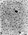
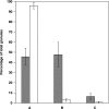
Piecemeal degranulation (PMD) has been recognized in two cases of human pheochromocytoma from the adrenal medulla, which were studied by transmission electron microscopy. Tumour pheochromocytes presented a highly characteristic cytoplasmic admixture of normal resting granules, swollen granules with eroded matrices and enlarged empty containers. Chromaffin granules that appeared to be normal and altered granules maintained their individual structure, and did not fuse with each other or with the plasma membrane. In accordance with the currently accepted model for granule discharge during PMD, electron-dense or clear vesicles 30-150 nm in diameter were seen either attached to the surface of chromaffin granules and the plasma membrane or free in the cytosol. This is the first description of PMD in human adrenal chromaffin cells and, in addition, is the first report of PMD in tumour secretory cells. These findings add further to the concept that PMD may have a broader spectrum of expression than hitherto recognized.
Figures


Similar articles
-
Ultrastructural morphology of adrenal chromaffin cells indicative of a process of piecemeal degranulation.Anat Rec A Discov Mol Cell Evol Biol. 2003 Feb;270(2):103-8. doi: 10.1002/ar.a.10013. Anat Rec A Discov Mol Cell Evol Biol. 2003. PMID: 12524685
-
Chromaffin cells in the adrenal homolog of Aphanius fasciatus (teleost fish) express piecemeal degranulation in response to osmotic stress: a hint for a conservative evolutionary process.Anat Rec A Discov Mol Cell Evol Biol. 2006 Oct;288(10):1077-86. doi: 10.1002/ar.a.20372. Anat Rec A Discov Mol Cell Evol Biol. 2006. PMID: 16964607
-
Chromaffin granules in the rat adrenal medulla release their secretory content in a particulate fashion.Anat Rec A Discov Mol Cell Evol Biol. 2004 Mar;277(1):204-8. doi: 10.1002/ar.a.20004. Anat Rec A Discov Mol Cell Evol Biol. 2004. PMID: 14983514
-
Piecemeal degranulation as a general secretory mechanism?Anat Rec A Discov Mol Cell Evol Biol. 2003 Sep;274(1):778-84. doi: 10.1002/ar.a.10095. Anat Rec A Discov Mol Cell Evol Biol. 2003. PMID: 12923888 Review.
-
Cell secretion mediated by granule-associated vesicle transport: a glimpse at evolution.Anat Rec (Hoboken). 2010 Jul;293(7):1115-24. doi: 10.1002/ar.21146. Anat Rec (Hoboken). 2010. PMID: 20340095 Review.
Cited by
-
Ultrastructural evidence of a vesicle-mediated mode of cell degranulation in chicken chromaffin cells during the late phase of embryonic development.J Anat. 2009 Mar;214(3):310-7. doi: 10.1111/j.1469-7580.2008.01032.x. J Anat. 2009. PMID: 19245498 Free PMC article.
-
Mast Cell Targeted Chimeric Toxin Can Be Developed as an Adjunctive Therapy in Colon Cancer Treatment.Toxins (Basel). 2016 Mar 11;8(3):71. doi: 10.3390/toxins8030071. Toxins (Basel). 2016. PMID: 26978404 Free PMC article.
-
Suggestive evidence of a vesicle-mediated mode of cell degranulation in chromaffin cells. A high-resolution scanning electron microscopy investigation.J Anat. 2010 Apr;216(4):518-24. doi: 10.1111/j.1469-7580.2009.01198.x. Epub 2010 Jan 28. J Anat. 2010. PMID: 20136671 Free PMC article.
References
-
- Bastiaensen E, De Block J, De Potter WP. Neuropeptide Y is localized together with enkephalins in adrenergic granules of bovine adrenal medulla. Neuroscience. 1988;25:679–686. - PubMed
-
- Brooks JC, Carmichael SW. Ultrastructural demonstration of exocytosis in intact and saponin-permeabilized cultured chromaffin cells. Am. J. Anat. 1987;178:85–89. - PubMed
-
- Carmichael SW, Brooks JC, Malhotra RK, Wakade TD, Wakade AR. Ultrastructural demonstration of exocytosis in the intact rat adrenal medulla. J. Electron Microsc. Technol. 1989;12:316–322. - PubMed
-
- Crivellato E, Ribatti D, Mallardi F, Beltrami CA. Granule changes of human and murine endocrine cells in the gastro-intestinal epithelia are characteristic of piecemeal degranulation. Anat. Rec. 2002;268:353–359. - PubMed
Publication types
MeSH terms
LinkOut - more resources
Full Text Sources
Medical

