. 2005 Jan;206(1):55-67.
doi: 10.1111/j.0021-8782.2005.00371.x.

Traversing the intact/fibrillated joint surface: a biomechanical interpretation

Affiliations

Traversing the intact/fibrillated joint surface: a biomechanical interpretation

Neil D Broom et al. J Anat. 2005 Jan.

Abstract

Cartilage taken from the osteoarthritic bovine patellae was used to investigate the progression of change in the collagenous architecture associated with the development of fibrillated lesions. Differential interference contrast optical microscopy using fully hydrated radial sections revealed a continuity in the alteration of the fibrillar architecture in the general matrix consistent with the progressive destructuring of a native radial arrangement of fibrils repeatedly interconnected in the transverse direction via a non-entwinement-based linking mechanism. This destructuring is shown to occur in the still intact regions adjacent to the disrupted lesion thus rendering them more vulnerable to radial rupture. Two contrasting modes of surface rupture were observed and these are explained in terms of the absence or presence of a skewed structural weakening of the intermediate zone. A mechanism of surface rupture initiation based on simple bi-layer theory is proposed to account for the intensification of surface ruptures observed in the intact regions on advancing towards the fibrillation front. Focusing specifically on the primary collagen architecture in the cartilage matrix, this study proposes a pathway of change from intact to overt disruption within a unified structural framework.


Figures
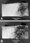
Fig. 1
Fig. 1
Cartilage-on-bone samples obtained from grade 1 (A) and grade 2 (B) bovine patellae showing the intact-fibrillated transition. In B note the increased density of cracks as the fibrillated region is approached.
Fig. 10
Fig. 10
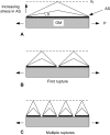
Schematic illustrating mode of rupture in a simple bi-layer model of cartilage in which there is structural bonding between two layers of contrasting strain-limiting properties. (Adapted from Broom et al. 1993.)
Fig. 2
Fig. 2
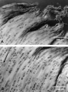
Micrographs showing zonal variation in matrix of intact region of a grade 1 patella adjacent to fibrillated region: (A) superficial zone; (B) intermediate or transition zone; (C) deep matrix. Arrows indicate radial direction.
Fig. 3
Fig. 3
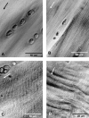
(A,B) Varying depths of penetration of radial propagating clefts in the intact region of a grade 2 patella. (C,D) Higher magnification views of the clefts in A and B, respectively. Arrows indicates radial direction.
Fig. 4
Fig. 4
(A) A partial delamination of the grade 2 articular surface without involving any tensile rupture in the plane of the surface. (B,C) Isolated curvilinear tears penetrating to different depths. (D) A more complex mode of delamination typical of the transition region. Arrows indicate radial direction.
Fig. 5
Fig. 5
(A) A distinct linear fibrous texture in mid and deep zones of grade 2 patella in the intact region close to the fibrillated region. (B) Higher resolution image of this same texture showing its finely crimped geometry (see small arrow). Long arrows indicate radial direction.
Fig. 6
Fig. 6
(A) Deeply clefted and distally flattened exposed matrix in severely fibrillated region of grade 2 patella. (B) Deeper matrix contiguous with A. The original articular surface is absent. Long arrow indicates radial direction; short arrow indicates cell cluster.
Fig. 7
Fig. 7
Micrographs showing progressive change in matrix texture with depth in the fibrillated region of a grade 2 patella. The faintly resolved radial texture of the deep zone (A) gives way to an increasingly visible radial texture and crimp as the exposed fibrillated surface is approached (B–D). Arrows indicate radial direction.
Fig. 8
Fig. 8
Transition region of a grade 2 patella with a near full thickness of the original cartilage but containing deep radial clefts. Short arrow indicates original articular surface. Long arrow shows radial direction.
Fig. 9
Fig. 9
Two-dimensional schema depicting progressive breakdown and re-arrangement of a network of fibrils in the general matrix based on a mediated, non-entwined mode of transverse linkage. Note how differing levels of elimination of the interconnecting elements lead to a range of destructured configurations.

References

    1. Bank RA, Soudry M, Maroudas A, Mizrahi J, Tekoppele JM. The increased swelling and instantaneous deformation of osteoarthritic cartilage is highly correlated with collagen degradation. Arthritis Rheumatism. 2000;43:2202–2210. - PubMed
    1. Bartels JE. Femoral–tibial osteoarthrosis in the bull: 1. Clinical survey and radiological interpretation. J. Am. Vet. Radiol. Soc. 1975;16:151–158.
    1. Broom ND. Further insights into the structural principles governing the function of articular cartilage. J. Anat. 1984a;139:275–294. - PMC - PubMed
    1. Broom ND. The altered biomechanical state of human femoral head osteoarthritic articular cartilage. Arthritis Rheumatism. 1984b;27:1028–1039. - PubMed
    1. Broom ND. The collagenous architecture of articular cartilage – a synthesis of ultrastructure and mechanical function. J. Rheumatol. 1986a;13:142–152. - PubMed

Publication types