

Infiltrative microgliosis: activation and long-distance migration of subependymal microglia following periventricular insults
- PMID: 15679892
- PMCID: PMC548677
- DOI: 10.1186/1742-2094-2-5
Infiltrative microgliosis: activation and long-distance migration of subependymal microglia following periventricular insults
Abstract
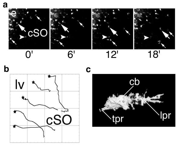
BACKGROUND: Subventricular microglia (SVMs) are positioned at the interface of the cerebrospinal fluid and brain parenchyma and may play a role in periventricular inflammatory reactions. However, SVMs have not been previously investigated in detail due to the lack of a specific methodology for their study exclusive of deeper parenchymal microglia. METHODS: We have developed and characterized a novel model for the investigation of subventricular microglial reactions in mice using intracerebroventricular (ICV) injection of high-dose rhodamine dyes. Dynamic studies using timelapse confocal microscopy in situ complemented the histopathological analysis. RESULTS: We demonstrate that high-dose ICV rhodamine dye injection resulted in selective uptake by the ependyma and ependymal death within hours. Phagocytosis of ependymal debris by activated SVMs was evident by 1d as demonstrated by the appearance of rhodamine-positive SVMs. In the absence of further manipulation, labelled SVMs remained in the subventricular space. However, these cells exhibited the ability to migrate several hundred microns into the parenchyma towards a deafferentation injury of the hippocampus. This "infiltrative microgliosis" was verified in situ using timelapse confocal microscopy. Finally, supporting the disease relevance of this event, the triad of ependymal cell death, SVM activation, and infiltrative microgliosis was recapitulated by a single ICV injection of HIV-1 tat protein. CONCLUSIONS: Subependymal microglia exhibit robust activation and migration in periventricular inflammatory responses. Further study of this population of microglia may provide insight into neurological diseases with tendencies to involve the ventricular system and periventricular tissues.
Figures


Similar articles
-
Neuroblast proliferation on the surface of the adult rat striatal wall after focal ependymal loss by intracerebroventricular injection of neuraminidase.J Comp Neurol. 2008 Apr 1;507(4):1571-87. doi: 10.1002/cne.21618. J Comp Neurol. 2008. PMID: 18236450
-
"Ependymal-in" Gradient of Thalamic Damage in Progressive Multiple Sclerosis.Ann Neurol. 2022 Oct;92(4):670-685. doi: 10.1002/ana.26448. Epub 2022 Jul 30. Ann Neurol. 2022. PMID: 35748636 Free PMC article.
-
Neuroinflammation induced by intracerebroventricular injection of microbial neuraminidase.Front Med (Lausanne). 2015 Mar 17;2:14. doi: 10.3389/fmed.2015.00014. eCollection 2015. Front Med (Lausanne). 2015. PMID: 25853134 Free PMC article.
-
Secondary Brain Injury Following Neonatal Intraventricular Hemorrhage: The Role of the Ciliated Ependyma.Front Pediatr. 2022 Jun 30;10:887606. doi: 10.3389/fped.2022.887606. eCollection 2022. Front Pediatr. 2022. PMID: 35844746 Free PMC article. Review.
-
Microgliosis in the Injured Brain: Infiltrating Cells and Reactive Microglia Both Play a Role.Neuroscientist. 2016 Apr;22(2):165-70. doi: 10.1177/1073858415572079. Epub 2015 Feb 11. Neuroscientist. 2016. PMID: 25672621 Review.
Cited by
-
Modulation of thalamic nociceptive processing after spinal cord injury through remote activation of thalamic microglia by cysteine cysteine chemokine ligand 21.J Neurosci. 2007 Aug 15;27(33):8893-902. doi: 10.1523/JNEUROSCI.2209-07.2007. J Neurosci. 2007. PMID: 17699671 Free PMC article.
-
Vivaria housing conditions expose sex differences in brain oxidation, microglial activation, and immune system states in aged hAPOE4 mice.Exp Brain Res. 2024 Mar;242(3):543-557. doi: 10.1007/s00221-023-06763-x. Epub 2024 Jan 11. Exp Brain Res. 2024. PMID: 38206365 Free PMC article.
-
Microglia activated by microbial neuraminidase contributes to ependymal cell death.Fluids Barriers CNS. 2021 Mar 23;18(1):15. doi: 10.1186/s12987-021-00249-0. Fluids Barriers CNS. 2021. PMID: 33757539 Free PMC article.
-
Mechanisms of chronic central neuropathic pain after spinal cord injury.Brain Res Rev. 2009 Apr;60(1):202-13. doi: 10.1016/j.brainresrev.2008.12.010. Epub 2008 Dec 25. Brain Res Rev. 2009. PMID: 19154757 Free PMC article. Review.
-
Interaction between Mycobacterium tuberculosis, Mycobacterium bovis, Mycobacterium avium subspecies paratuberculosis with the enteric glia and microglial cells.Gut Pathog. 2011 Dec 9;3:19. doi: 10.1186/1757-4749-3-19. Gut Pathog. 2011. PMID: 22151930 Free PMC article.
References
-
- Becher B, Prat A, Antel JP. Brain-immune connection: immunoregulatory properties of CNS-resident cells. Glia. 2000;29:293–304. - PubMed
-
- Ling E A, Kaur C, Lu J. Origin, nature and some functional considerations of intraventricular macrophages, with special reference to the epiplexus cells. Microsc Res Tech. 1998;41:43–56. - PubMed
Grants and funding
LinkOut - more resources
Full Text Sources
