. 2004 Oct 25;1(1):3.
doi: 10.1186/1742-9994-1-3.

Secondary neurons are arrested in an immature state by formation of epithelial vesicles during neurogenesis of the spider Cupiennius salei

Affiliations

Secondary neurons are arrested in an immature state by formation of epithelial vesicles during neurogenesis of the spider Cupiennius salei

Angelika Stollewerk. Front Zool. .

Abstract

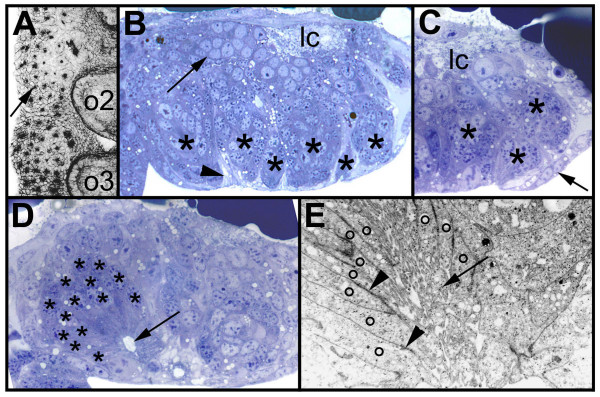
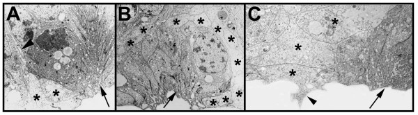
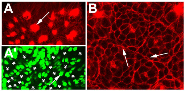
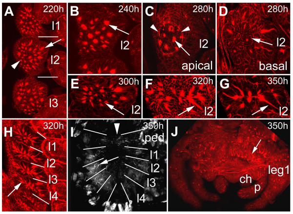
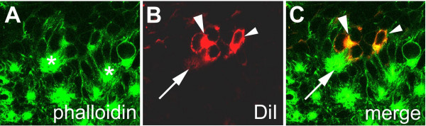
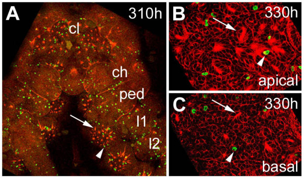
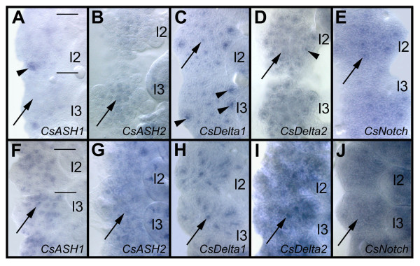
BACKGROUND: In the spider Cupiennius salei about 30 groups of neural precursors are generated per hemi-segment during early neurogenesis. Analysis of the ventral neuromeres after invagination of the primary neural precursor groups revealed that secondary neural precursors arise during late embryogenesis that partially do not differentiate until larval stages. RESULTS: In contrast to the primary groups, the secondary invaginating cells do not detach from each other after invagination but maintain their epithelial character and form so-called epithelial vesicles. As revealed by dye labeling, secondary neural precursors within epithelial vesicles do not show any morphological features of differentiation indicating that the formation of epithelial vesicles after invagination leads to a delay in the differentiation of the corresponding neural precursors. About half of the secondary neural precursor groups do not dissociate from each other during embryogenesis indicating that they provide neural precursors for larval and adult stages. CONCLUSIONS: Secondary neural precursors are arrested in an immature state by formation of epithelial vesicles. This mechanism facilitates the production of larval neural precursors during embryogenesis. I discuss the evolutionary changes that have occured during neural precursor formation in the arthropod group and present a model for the basal mode of neurogenesis.


Figures

Figure 1
Figure 1
(A-E): Morphology of the secondary invagination sites. Confocal micrograph (A, inverted) of a flat preparation of an embryo stained with phalloidin-rhodamine and light micrographs (B-D) and electron micrograph (E) of transverse sections through prosomal hemi-neuromeres. The midline is to the right. (A) Final pattern of the primary invagination sites in the opisthosomal segments 1 and 2. The invagination sites are arranged in 7 rows. The black dots correspond to the constricted cell processes of the individual precursor groups that are attached to the apical surface (arrow). (B) Morphology of the secondary invagination sites. At 250 hours the secondary invaginating cell groups (asterisks) are still attached to the apical surface. The individual groups are isolated by brighter sheath cells (arrowhead). The primary precursor groups have dissociated (arrow) and form basal cell layers. The longitudinal connective (lc) is already visible at the basal side. (C) The secondary invagination sites (asterisks) loose contact to the apical surface, when the epidermis (arrow) overgrows the ventral neuromeres. (D) After invagination the secondary neural precursors (asterisks) remain attached to each other forming epithelial vesicles. The cell processes run parallel to each other and extend to a lumen (arrow). (E) The cell processes (o) of the invaginating cells of a group are opposed to each other and the lumen between the cell processes is filled with microvilli (arrow). Cell junctions connect the individual processes (arrowheads). lc, longitudinal connective; o2 to o3, opisthosomal hemi-segments 2 to 3.
Figure 2
Figure 2
(A-C): Secondary invagination sites are surrounded by sheath cells. Electron micrographs of transverse sections through prosomal hemi-neuromeres. (A,B) Invagination sites (arrows) are surrounded by sheath cells (asterisks) that appear translucent in the electron microscope. The sheath cells extend processes (arrowhead) that enwrap the individual invagination sites. (C) Sheath cells that are located in the apical cell layer form bizarre shapes that extend into the cell free space at the ventral side of the embryo (arrowhead). The sheath cells are labeled with asterisks, the arrow points to an invagination site.
Figure 3
Figure 3
(A-B): The nuclei of cells within the secondary invagination sites are located basally. Confocal micrographs of flat preparations of embryos double-stained with phalloidin-rhodamine (red) and YOYO (green) (A,A') and single stained with phalloidin-rhodamine (B). (A,A') The apical optical section at 250 hours of development shows that the secondary invagination sites (arrow) are still attached to the apical surface. The nuclei of the secondary precursors are located basally, as revealed by the absence of nuclei staining in the apical cell layer. The asterisks in A' indicate the positions of the cell processes of the secondary invagination sites (compare to A). (B) The basal optical section shows the distinct morphology of the sheath cells (arrows) that subdivide the individual invagination sites.
Figure 4
Figure 4
(A-j): Invagination of secondary neural precursors and formation of epithelial vesicles. (A-F) Confocal micrographs of flat preparations of embryos stained with phalloidin-rhodamine. (B-G) Flat preparations of the fourth prosomal hemi-segments. (A) At 220 hours about 25 secondary invagination sites form (arrow). There is no clear dividing line between the formation of secondary invagination sites (arrow) and invagination of primary neural precursors. Some primary invagination sites are still visible (arrowhead) The bars indicate the segment borders. (B) Apical optical section of the pattern of secondary invagination sites (arrow) at 240 hours of development. (C) Epidermal cells overgrow the ventral neuromeres between 250 and 300 hours (arrowheads) The arrow points to a secondary invagination group. (D) After invagination the individual cells of a groups remain attached to each other forming epithelial vesicles (arrow). (E) At 300 hours the anterior-posterior extension of the individual hemi-segments has been reduced leading to a rearrangement in the positions of the invaginated cell groups (arrow). (F) After 320 hours 8 of the 25 invaginated cell groups are no longer visible indicating that the cells have detached from each other. The arrow points to an invaginated cell group. (G) 10 cell groups are still visible at hatching (arrow). (H) Overview of the arrangement of epithelial vesicles (arrow) of the four prosomal hemi-segments corresponding to the four walking legs. The anterior-posterior reduction in size is clearly visible (compare to A). The bars indicate the segment borders. (I) Flat preparation of the prosoma at hatching. Epithelial vesicles are still visible (arrow). The bars indicate the segment borders, the arrowhead points to the midline. (J) Flat preparation of the brain at 350 hours. The arrow points to epithelial vesicles. ch, chelicera; l1 to l4, prosomal neuromeres corresponding to walking leg 1 to 4; leg 1, walking leg 1.p, pedipalp; ped, pedipalpal neuromere.
Figure 5
Figure 5
(A-C): DiI-labeling of cells within epithelial vesicles. Flat preparation of the fourth prosomal hemi-neuromere of an embryo labeled with DiI (red) and stained with phalloidin-rhodamine (green). (A-C) Invaginated cells in 40 segments of 10 embryos were labelled with DiI (red) and stained with phalloidin-FITC (green). The cells of a group (A, asterisks) are attached to each other (B,C large arrow head) and their short, thin cell processes run parallel to each other (B,C arrow). They do not show any morphological features of differentiation, i.e. they do not grow long thin dendritic or axonal processes. The small arrows (B,C) point to a cell of an adjacent invagination group.
Figure 6
Figure 6
(A-C): Mitotic pattern in the ventral neuromeres after formation of the secondary invagination sites. Flat preparations of embryos stained with phalloidin-rhodamine (red) and anti-Phospho-Histon 3 (green). (A) Only scattered mitotic cells (arrowhead) are present in the ventral neuromeres after invagination of the secondary neural precursors (arrow). The pattern of cell divisions in the cephalic lobe and the prosomal segments at 310 hours of development is representative for the late embryonic stages. (B) Optical section through apical cell layers of the fourth prosomal hemi-neuromere. Only a few mitotic cells (arrowhead) are associated with epithelial vesicles. (C) A similar pattern is visible in basal cell layers of the same neuromere. The arrowhead points to a dividing cell, the arrow points to a dissociating epithelial vesicle. ch, cheliceral neuromere; cl, cephalic lobe; l1 to l2, prosomal hemi-neuromeres corresponding to walking legs 1 to 2; ped, pedipalpal hemineuromere.
Figure 7
Figure 7
(A-J): Proneural and neurogenic genes are re-expressed during formation of the secondary neural precursors. Flat preparations of the fourth and fifth prosomal hemi-segments after in situ hybridisation of whole embroys. (A-E) 220 hours of development, (F-J) 250 hours of development. Anterior is at the top, the midline to the left. (A) At 220 hours, CsASH1 expression has been down-regulated in all primary neural precursors (arrow) with the exception of one group (arrowhead). (B) At this time the pan-neural gene CsASH2 is still weakly expressed in the primary neural precursors (arrow). (C) CsDelta transcripts accumulate in the secondary invagination sites (arrow heads), while transcripts are down-regulated in the primary precursor groups. (D) A similar expression, although weaker, is visible after CsDelta2 in situ hybridisation. The arrow points to a region where CsDelta2 has been down-regulated, the arrowhead indicates expression in the secondary neural precursors. (E) CsNotch remains expressed at low levels in the ventral neuroectoderm. An up-regulation of CsNotch transcripts is visible in the secondary invagination groups (arrow). (F) At 250 hours CsASH1 expression can be detected in the secondary invagination sites (arrow), although it is not expressed in all of them. (G) CsASH2 seems to be expressed weakly in all secondary invaginating cells groups (arrow). (H) A high accumulation of CsDelta1 transcripts is visible in about 10 of the invagination sites (arrow), (I) while CsDelta2 seems to be xpressed in all invagination groups (arrow). (J) Cs Notch transcripts can be detected in all neuroectodermal cells at this time. l2 to l3, walking leg 2 to 3.

Similar articles

Cited by

References

    1. Stollewerk A, Weller M, Tautz D. Neurogenesis in the spider Cupiennius salei. Development. 2001;128:2673–2688. - PubMed
    1. Stollewerk A. Recruitment of cell groups through Delta/Notch signalling during spider neurogenesis. Development. 2002;129:5339–5348. doi: 10.1242/dev.00109. - DOI - PubMed
    1. Dove H, Stollewerk A. Comparative analysis of neurogenesis in the myriapod Glomeris marginata (Diplopoda) suggests more similarities to chelicerates than to insects. Development. 2003;130:2161–2171. doi: 10.1242/dev.00442. - DOI - PubMed
    1. Kadner D, Stollewerk A. Neurogenesis in the chilopod Lithobius forficatus suggests more similarities to chelicerates than to insects. Dev Genes Evol. 2004;214:367–379. doi: 10.1007/s00427-004-0419-z. - DOI - PubMed
    1. Mittmann B. Early neurogenesis in the horseshoe crab and its implication for arthropod relationships. Bio Bull. 2002;203:221–222. - PubMed