. 2005 Feb;79(4):2221-9.
doi: 10.1128/JVI.79.4.2221-2229.2005.

RNA recombination of hepatitis delta virus in natural mixed-genotype infection and transfected cultured cells

Affiliations

RNA recombination of hepatitis delta virus in natural mixed-genotype infection and transfected cultured cells

Tzu-Chi Wang et al. J Virol. 2005 Feb.

Abstract

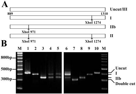
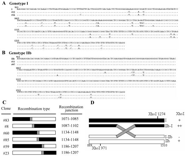
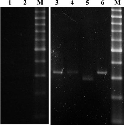
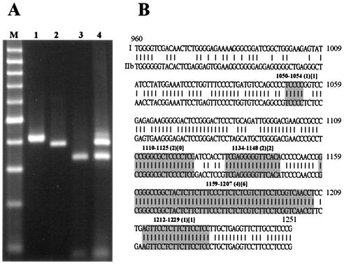
Most RNA viruses encode their own RNA polymerases for genome replication, and increasing numbers of them appear to be capable of undergoing RNA recombination. Here, we provide the first report of intergenotypic recombination in hepatitis delta virus (HDV), the only animal RNA virus that requires host RNA polymerase(s) for viral replication. In vivo, we analyzed RNA species derived from the serum of a patient with mixed genotype I and genotype IIb HDV infection by using multiple restriction fragment length polymorphism (RFLP) assays and sequence analysis of cloned reverse transcription (RT)-PCR products. Six HDV recombinants were isolated from 101 tested clones, and HDV recombination in this patient was further confirmed by RT-PCR with genotype-specific primer pairs. Analysis of the recombination junctions suggested that the HDV genome rearrangement occurred through faithful homologous recombination. We then used an RNA cotransfection cell culture system to investigate HDV RNA recombination in vitro. We found that HDV recombinants could indeed be detected in the transfected cells; some of these possessed recombination junctions identical to those identified in vivo. Furthermore, using a PCR-independent RNase protection assay, we were able to readily identify the recombined HDV RNA species in cultured cells. Taken together, our results demonstrate that HDV RNA recombination occurs in both natural HDV infections and cultured cells, thereby presenting a novel mechanism for HDV evolution.


Figures

FIG. 1.
FIG. 1.
XhoI RFLP analysis for detection of different HDV genotypes in a mixed infection. (A) XhoI restriction sites in the amplified sequences of different genotypes. The PCR products of genotypes I and IIb each have a single XhoI site, at nt 1274 and 971, respectively. Genotype II PCR products contain both of these XhoI sites, whereas genotype III contains no XhoI site in this region. (B) The XhoI-cleaved PCR products of HDV genomes were electrophoresed on a 3% agarose gel and stained with ethidium bromide. Lanes: M, 100-bp-ladder molecular size markers; 1 and 10, undigested PCR products; 2 and 9, digested genotype I PCR products; 3 and 8, digested genotype IIb PCR products; 4, digested PCR products of mixed in vitro-transcribed genotype I and IIb RNAs; 5, digested PCR products of mixed total cellular RNAs extracted from cells transfected separately with genotype I and IIb RNAs; 6, a complex RFLP pattern obtained from the HDV patient; 7, digested genotype II PCR products.
FIG. 2.
FIG. 2.
HDV-related sequences detected in the serum of a patient with mixed genotype I and IIb infection. (A and B) Alignment of nucleotide sequences (nt 913 to 1280) from representative HDV isolates of genotypes I (A) and IIb (B), and sequences identified in the mixed infection of HDV. Dots indicate conserved nucleotides. Sources of the representative isolates of different genotypes are as follows: I, HDV genotype I isolated from Italy (GenBank accession number M21012) (28); IIb, genotype IIb from the Taiwan-IIb-1 clone (GenBank accession number AF209859) (60). The sequences obtained from two clones each of HDV genotypes I (clones 12 and 16) and IIb (clones 11 and 97) from the mixed infection are shown. (C) Schematic demonstration of HDV recombinants identified by sequence analysis. The recombination junctions are shaded in gray and summarized on the right. The genotype I and IIb sequences are indicated by closed and open bars, respectively. (D) Restriction maps of the potential clones carrying recombinant HDV genomes. The XhoI RFLP profiles and the predicted genome organization of recombinants 5′-I-IIb-3′ and 5′-IIb-I-3′ are summarized on the right.
FIG. 3.
FIG. 3.
Detection of HDV recombinants by use of genotype-specific primers. cDNAs generated with genotype I- or IIb-specific primers served as the templates for subsequent PCRs with recombinant-specific primer pairs. The PCR products were cleaved with XhoI, separated on 3% agarose gels, and stained with ethidium bromide. The observed 5′-IIb-I-3′ recombinant contains one XhoI site (nt 971), whereas the 5′-I-IIb-3′ recombinant lacks a XhoI site. Lanes: M, 100-bp-ladder molecular size markers (the dominant band is 500 bp in size); 1 and 2, undigested PCR products of mixture of in vitro-transcribed genotype I and IIb RNA by use of primer pairs I5′-1-IIb3′-1 and IIb5′-1-I3′-1, respectively; 3 and 6, undigested PCR products obtained from the HDV patient with primer pairs I5′-1-IIb3′-1 and IIb5′-1-I3′-2, respectively; 4 and 5, digested PCR products obtained from the HDV patient with primer pairs I5′-1-IIb3′-1 and IIb5′-1-I3′-2, respectively.
FIG. 4.
FIG. 4.
Primary structure of the crossover regions of intergenotypic HDV recombinants identified in a natural mixed-genotype HDV infection. Genomic sequences (nt 952 to 1280) of HDV genotypes I and IIb are given. Short lines depict the homologous bases between two genotypes. The crossover regions identified using genotype-specific primers are indicated by gray shading, while those obtained using consensus primers are indicated by rectangles.
FIG. 5.
FIG. 5.
HDV recombination in transfected cultured cells. (A) XhoI RFLP analysis of PCR products amplified from total RNA extracted from cotransfected cultured cells by use of consensus primers. Lanes: M, 100-bp-ladder molecular size markers (the dominant band is 500 bp in size); 1, undigested PCR products; 2, digested genotype I PCR products; 3, digested genotype IIb PCR products; 4, a complex RFLP pattern obtained from transfected cultured cells. (B) Primary structure of the crossover regions of intergenotypic HDV recombinants identified in transfected cultured cells by use of genotype-specific primers. The nucleotide sequences of the genomic segments (nt 960 to 1251) of HDV genotypes I (Italian clone) and IIb (Taiwan-IIb-1 clone) are given. Short lines depict the homologous bases between the two genotypes. The crossover regions identified are indicated by gray shading. Numbers given in parentheses and square brackets indicate the number of 5′-I-IIb-3′ and 5′-IIb-I-3′ recombinant clones, respectively.
FIG. 6.
FIG. 6.
Detection of HDV RNA recombinants with an RNase protection assay. Probes specific to the HDV RNA recombinant with the crossover region located at nt 1159 to 1207 were synthesized in vitro, hybridized with various RNA samples, and subjected to RNase digestion for detection of the HDV RNA recombinants. Lanes 7 to 12 represent the results of a longer exposure of the data shown in lanes 1 to 6. Lanes: 1 and 7, no-target, no-RNase control lanes corresponding to the full-length probe; 2 and 8, probe hybridized with a complementary in vitro-transcribed HDV RNA; 3 and 9, probe hybridized to RNA samples extracted from cotransfected cultured cells; 4 and 10, probe hybridized to RNA samples extracted from untransfected cells; 5 and 11, probe hybridized to mixtures of in vitro-transcribed genotype I and IIb RNAs; 6 and 12, probe hybridized to mixtures of total RNA extracted from cells independently transfected with genotype I and IIb HDV RNAs. The probe and protected bands are schematically depicted on the right. The genotype I, genotype IIb, and vector sequences are indicated by a closed box, open box, and thin line, respectively.

Similar articles

Cited by

References

    1. Ambros, S., J. C. Desvignes, G. Llacer, and R. Flores. 1995. Pear blister canker viroid: sequence variability and causal role in pear blister canker disease. J. Gen. Virol. 76:2625-2629. - PubMed
    1. Branch, A. D., B. J. Benenfeld, B. M. Baroudy, F. V. Wells, J. L. Gerin, and H. D. Robertson. 1989. An ultraviolet-sensitive RNA structural element in a viroid-like domain of the hepatitis delta virus. Science 243:649-652. - PubMed
    1. Brazas, R., and D. Ganem. 1996. A cellular homolog of hepatitis delta antigen: implications for viral replication and evolution. Science 274:90-94. - PubMed
    1. Cascone, P. J., C. D. Carpenter, X. H. Li, and A. E. Simon. 1990. Recombination between satellite RNAs of turnip crinkle virus. EMBO J. 9:1709-1715. - PMC - PubMed
    1. Casey, J. L., T. L. Brown, E. J. Colan, F. S. Wignall, and J. L. Gerin. 1993. A genotype of hepatitis D virus that occurs in northern South America. Proc. Natl. Acad. Sci. USA 90:9016-9020. - PMC - PubMed

Publication types